

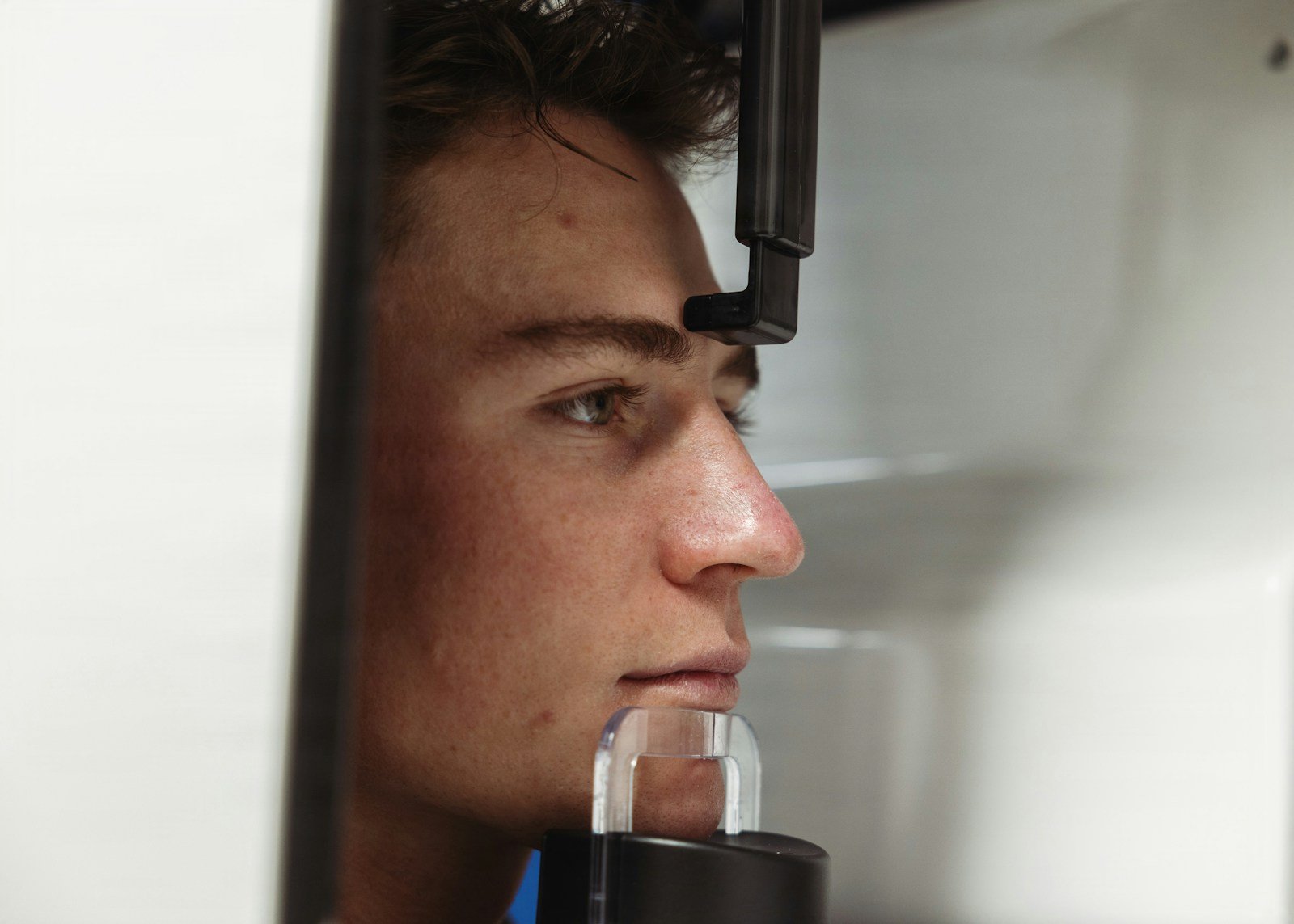
Somewhere between a child’s twelfth and fourteenth birthday, the lower jaw does most of its growing — a surge that can last barely a year. Catch it, and a brace or a growth-guiding appliance can coax a developing bite into place almost effortlessly. Miss it, and the same correction may later mean surgery. For decades, orthodontists have tried to spot that window by reading cervical vertebral maturation — the shape of three small bones in the neck — on a side-view X-ray. It is, by every honest account, a bit of a guess.
The 30-Second Version
- A 2025 PLOS One study trained AI to read cervical vertebral maturation (CVM) — the neck-bone shorthand for a teenager’s growth stage — from 3,600 orthodontic X-rays across six centers.
- Two strategies were tested head-to-head: eyeballing the bone shapes (qualitative) versus measuring them (quantitative). The measuring route won, 78% accuracy to 71%.
- The AI placed its measurement landmarks within 1 mm of human experts 97% of the time, and staged a batch of scans in 7 minutes that took a junior orthodontist close to an hour.
- But roughly 1 in 5 scans was still mis-staged — and the model was shakiest in the middle stages, the exact window the growth spurt hides in.
It sounds like the end of a decades-old headache. But the details are more interesting than the headline. The study, published in PLOS One in May 2025 by Abbas Ahmed Abdulqader and colleagues across six orthodontic centers in China, is the first to put the two ways of automating this read side by side and ask a sharper question than “can AI do it?” — namely, which way of doing it actually helps, and where does it still break?
The study, in one glance
The team started with 6,000 lateral cephalograms — the standard side-view head X-ray of orthodontics — and filtered down to 3,600, deliberately balanced so each of the six cervical stages (CS1 through CS6) was equally represented. They split the images 8:1:1 into training, tuning, and a 360-scan test set. One pipeline used a ResNet-101 network to judge the shape of the C2–C4 vertebrae (the qualitative method). The other used a YOLOv3 network to pin down 13 anatomical landmarks, measured concavities and height ratios from them, then let a CatBoost classifier assign the stage (the quantitative, or QCVM, method). Both were graded against staging by three orthodontists, whose own agreement was good but not perfect (Cohen’s kappa 0.78–0.85).
Where AI cervical vertebral maturation reads help
The genuine win here is not raw cleverness; it is consistency and speed. Growth staging has always been bedevilled by subjectivity — two clinicians can look at the same neck bones and disagree, and the same clinician can disagree with themselves on a different day. The quantitative pipeline sidesteps some of that by turning the judgement into numbers: when it measured the vertebrae, its figures tracked the orthodontists’ own measurements almost exactly (Pearson correlation up to 0.98, with tiny error). And it is fast. The AI staged all 360 test scans in about seven minutes; placing the measurement landmarks by hand took the junior orthodontists nearly six hours for the same set. In a busy clinic, a consistent, instant second read — one that can also place cephalometric landmarks without a human tracing every point — is a real convenience, and because a cervical reading can stand in for a separate hand-and-wrist X-ray, it can even spare a growing child some radiation.
But how often is it simply wrong?
About one time in five. The best method reached 78.3% accuracy; the shape-reading method, 71.1%. That is useful, but it is nowhere near a settled verdict — and the errors are not spread evenly. Both methods were strongest at the extremes, where the bones look obviously immature (CS1) or obviously finished (CS6), and weakest in the crowded middle. In the shape-reading model, stage CS2 scraped a precision of just 0.54 and CS3 a recall of only 0.56.
⚠ The error lands where it hurts most
The classic orthodontic growth spurt — the burst of jaw growth clinicians most want to catch — falls between the middle stages, CS3 and CS4. That is precisely where both AI methods were least reliable. A model that nails the easy start and finish but blurs the pivotal middle is weakest at the one judgement the whole exercise exists to make.
It’s a weather app that’s spot-on about last week and next month, but vague about tomorrow — the one forecast you were actually going to plan around.
There is a deeper caveat in how the test was run. The 360 test scans came from the same six-center pool as the training data, split at random, so this is an internal check, not an independent, external validation on scans from a new clinic or population. The “gold standard” the AI was scored against was the orthodontists’ own staging — itself subjective — and the authors note they did not cross-check it against hand-wrist radiographs or chronological age. One of the authors is affiliated with a commercial AI company, and all the data came from a single country. None of that makes the work wrong; it makes it a promising proof of concept rather than a validated clinical tool.
If the AI and the orthodontist disagree, who’s right?
This is the quiet trap in any model trained to imitate experts. The AI learned the stages from human staging, so at its very best it can only match the clinicians who taught it — it cannot be more correct than they were, and it inherits their disagreements. When three orthodontists only agreed with each other at a kappa of 0.78–0.85, a machine trained on their labels is not resolving the ambiguity of growth staging; it is reproducing it, faster. That is worth having, but it is not the same as truth.
Who might this leave out?
Every image in this study came from one population, and cervical vertebrae — like faces and teeth — vary across people. A model tuned to one group’s anatomy can quietly misread another’s until it is tested there, which this one has not been. Access cuts the same way: the children most likely to benefit from well-timed, growth-guided orthodontics are often the least likely to have a cephalogram taken in the first place. A tool that makes expert reading faster helps most the clinics that already have the experts and the imaging; it does nothing, on its own, for the places that have neither.
What this means for you
If you’re a patient or parent
If your orthodontist mentions an AI growth-stage read, treat it as one input, not a ruling. Timing a child’s treatment draws on several signs — height changes, erupting teeth, the X-ray — and no single stage number should rush you into, or hold you back from, starting treatment. A good clinician uses it to confirm a hunch, not to replace the conversation.
If you’re a clinician
The quantitative pipeline is appealing for consistency and record-keeping, but be most skeptical of a single-stage output around CS3–CS4, where accuracy sagged. Corroborate with other maturity indicators, and ask for external validation on a population like yours before you let a stage call drive a treatment-timing decision.
The bottom line
AI can make the growth read faster and steadier, and the measuring route is clearly the smarter of the two. But staging a child’s maturation is ultimately a prediction about the future, and no neck X-ray — read by a human or a machine — sees the future perfectly. This is a sharper second opinion on timing, delivered in minutes. It is an assistant to the clinician’s judgement, not an oracle that replaces it.
Frequently asked questions
What is cervical vertebral maturation (CVM)?
It’s a way of estimating a growing person’s skeletal maturity — roughly, how much growing they have left — from the shape of the second, third, and fourth bones of the neck as they appear on a standard side-view orthodontic X-ray (a lateral cephalogram). Orthodontists use it to time treatments that work best during active growth, and it can serve as an alternative to a separate hand-and-wrist X-ray.
How accurate was the AI in this study?
The quantitative method, which measured the bones and then classified the stage, reached 78.3% accuracy on the 360-scan test set. The qualitative method, which judged bone shape directly, reached 71.1%. Both were most accurate at the earliest and latest stages and least accurate through the middle stages (CS2–CS4). According to PubMed, this is reported in the study (DOI: 10.1371/journal.pone.0323776).
Can AI decide when to start my child’s orthodontic treatment?
No. The AI estimates a growth stage; it doesn’t make treatment decisions. Those depend on the clinical picture as a whole, and the model was weakest in exactly the middle stages where growth-timing calls are most delicate. It’s best used as a fast, consistent second read for an orthodontist, not a substitute for one.
Is this ready to use in clinics today?
Not yet. The results come from an internal test on scans from the same six centers that supplied the training data, with no external validation on a new clinic or population, no cross-check against other maturity measures, and data from a single country. It’s a strong proof of concept that needs independent testing before routine clinical use.
Does AI growth staging mean fewer X-rays?
Potentially. Because a cervical reading from a cephalogram many orthodontic patients already have can substitute for a dedicated hand-and-wrist radiograph, automating it could reduce one source of imaging — though the cephalogram itself still involves a small dose.
Source & author credit
This article interprets, and does not reproduce, the following peer-reviewed study. All figures are the authors’ original findings. According to PubMed:
Abdulqader AA, Jiang F, Almaqrami BS, Cheng F, Yu J, Qiu Y, Li J. Deep learning approaches for quantitative and qualitative assessment of cervical vertebral maturation staging systems. PLoS One. 2025;20(5):e0323776. DOI: 10.1371/journal.pone.0323776
ORCID — Juan Li: 0000-0001-8320-2406
Published open access under a Creative Commons Attribution 4.0 (CC BY 4.0) license. Decadentry is an independent educational publication and is not affiliated with the study’s authors. Every article is checked against its primary source under our editorial standards; see more orthodontics research.

Leave a Reply