
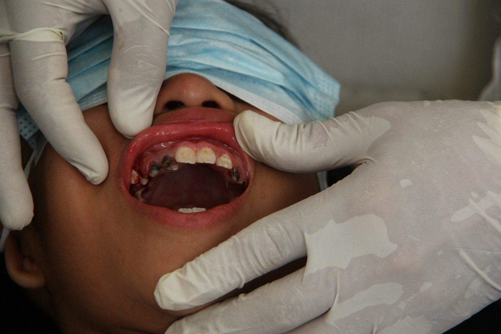
In a community hall in eastern Taiwan, a health worker with no specialist training lifts a phone to a resident’s open mouth, takes a plain white-light photo, and an app paints a colored box on the screen: green, yellow, or red. Red means see a specialist — this could be cancer. For a disease that still kills roughly half the people it strikes within five years, that little colored box is carrying a lot of weight. This is mobile AI oral cancer screening as it actually happens in the field — not a tidy benchmark, but a real screening day with bad lighting, nervous patients, and a camera held by someone who isn’t a dentist.
The 30-Second Version
- A fixed, pre-trained AI triage app was tested live in community oral-cancer screenings in eastern Taiwan — 602 residents, 4,283 usable mouth photos taken by non-specialists on handheld devices.
- For its high-risk “red” flag it caught 75% of lesions experts later judged dangerous, and ranked risk well (AUROC 0.957).
- The catch: only 17.6% of its red flags were real — about five in six were false alarms — and it still missed 4 of 16 truly high-risk lesions.
- Honest caveat: the “gold standard” was a panel of specialists viewing the same photos (not biopsies), and they could see the AI’s answers — so the scores likely flatter the tool.
It sounds like exactly the tool underserved communities need. But look closer. The study — Liu and colleagues, published in JMIR mHealth and uHealth in 2026 — is unusual precisely because it refused to grade itself in a lab. Most dental-AI papers report how a model scores on a curated image set; unlike the lab-benchmarked oral-cancer cytology test we covered earlier, which hit near-perfect AUROC on clean brushed-cell samples, this one asked a harder question: what happens when you hand a locked, already-trained system to ordinary screeners and turn it loose on real mouths? The answer is more sobering — and more useful — than another near-perfect accuracy figure.
The study, in one glance
This was a prospective field evaluation — the researchers watched the system work in real time rather than replaying old data. From June to December 2025, trained non-specialist staff photographed the mouths of 602 community residents during routine screenings. An on-device quality check rejected blurry or badly lit shots on the spot — 533 of 4,816 images (about 11%) were thrown out — leaving 4,283 usable photos. The AI sorted each finding into green (nothing to act on), yellow (follow up) or red (refer to a specialist now). Three board-certified oral and maxillofacial specialists then reviewed every image and, by consensus, decided what the right call should have been.
Where mobile AI oral cancer screening genuinely helps
Start with what impressed. The system’s discriminative power was strong: AUROC was 0.957 for red, 0.931 for yellow and 0.964 for green — meaning that, handed a random high-risk lesion and a random low-risk one, its confidence score ranked them correctly about 96% of the time. The yellow “follow-up” tier was both sensitive (92.7%) and reasonably precise (81.4%), and the red flag caught three of every four lesions the experts judged dangerous. In a region where oral cancer is common — betel-quid chewing is a major driver — and specialists are scarce, a cheap phone-based first pass that reliably says “look here” could shorten the distance between a suspicious patch and an expert’s eyes. Triage was never meant to diagnose; its job is to decide who jumps the queue.
But what does a red flag actually mean?
Here is the uncomfortable number. The AI raised 68 red flags. After expert review, just 12 were truly high-risk — a positive predictive value of 17.6%. Put plainly: roughly five of every six “urgent” alarms were false. That isn’t necessarily a broken model. In community screening, dangerous lesions are rare — Taiwanese programs put the prevalence of referral-worthy lesions at about 0.9% to 2.2% — and when a condition is that rare, even a specific test produces mostly false positives. This is arithmetic, not a malfunction. But arithmetic has consequences for the person on the other end of a red box.
⚠ A red flag is a reason to look, not a reason to panic
At 17.6% precision, a red result should trigger a specialist visit — not a diagnosis, and not a frightening conversation. The authors are blunt that this is “human-supervised referral-prioritization support,” not autonomous diagnosis. A patient told “the AI flagged something” who hears “I have cancer” has been failed by the framing, not the finding.
Think of it less like a cancer test and more like a smoke alarm in a big building: it shrieks at burnt toast far more often than at a real fire — but you still want it there, because the one time it catches the fire, it matters.
And the misses matter as much as the false alarms. The model missed 4 of 16 genuinely high-risk lesions — two it failed to detect at all, two it filed as lower-risk. The culprits were exactly the hard cases: lesions near the edge of the frame, mixed red-and-white patches, low contrast. A tool that stumbles on subtle, partially captured lesions is weakest in precisely the situation where a careful human second look matters most.
Can we trust the scorecard itself?
This is where an honest reader slows down. There was no biopsy. The “truth” the AI was graded against was a consensus of three specialists looking at the same photographs — a sensible operational standard for a screening study, but not a tissue diagnosis. And in a detail that inflates the AI’s numbers, the reviewers could see its triage labels as they judged; the authors concede an unblinded reference standard “may have overestimated agreement.” With only 16 true high-risk lesions in the entire study, the 75% sensitivity comes wrapped in a wide confidence interval (50.5–89.8%) — the real figure could be little better than a coin toss or close to perfect. None of this is fatal; it’s the honest ceiling on a single-region pilot, and it sits alongside the rest of our diagnostics and imaging coverage, where “promising but unvalidated” is the rule, not the exception.
Who is this really for — and who gets left out?
The whole pitch here is equity: mobile, cheap, operable by non-specialists, aimed at the rural and high-risk populations that specialist clinics never reach. That is the strongest argument for the approach — but a field tool is only as good as the field. The system discarded 11% of images for quality and analyzed only the ones that passed, so the real-world burden of re-takes, and of mouths that never yield a usable photo, is understated. It was tested in one region, on one population, with one set of devices and operators. Whether it holds up on a different phone, a different clinic, or a different population’s mucosa — darker pigmentation, different lesion patterns — is simply unknown. Equity is the promise; proving it requires the multi-center work the authors say still has to be done.
What this means for you
If you’re a patient
If a screening app flags your mouth, take it seriously enough to see a dentist or specialist — but don’t read a red box as a diagnosis. It’s a prompt to get a proper exam, where a clinician looks, feels, and if needed, biopsies. A green result isn’t a guarantee either: keep your regular checkups, especially if you smoke, drink heavily, or chew betel quid.
If you’re a clinician
Treat these systems as a queue-sorter, not a colleague. The value is in prioritizing referrals where specialists are scarce, not in replacing your eyes — and never in reassuring a patient. Budget for the false-positive load (it creates real anxiety and workload), demand blinded, biopsy-anchored, multi-center validation before trusting the numbers, and remember the tool is weakest on the subtle, edge-of-frame lesions you’re there to catch.
The bottom line
The most honest thing about this study is what it refused to claim. It didn’t say the AI diagnoses oral cancer; it said the AI can help decide who to look at next, under a specialist’s supervision, in a place where that help is genuinely scarce. That’s a smaller promise than the headlines want — and a more trustworthy one. The future of screening in underserved communities may well be a phone in a non-specialist’s hand. But the phone’s job is to point; the deciding still belongs to a human.
Frequently asked questions
Does this AI diagnose oral cancer?
No. It triages — sorting mouths into green, yellow and red to decide who needs a specialist. Diagnosis still requires a clinical exam and, when warranted, a biopsy. The study explicitly frames the tool as human-supervised referral support, not autonomous diagnosis.
If it catches 75% of dangerous lesions, why were only 17.6% of its red flags real?
Those measure different things. Sensitivity (75%) is how many true high-risk lesions it caught. Precision, or positive predictive value (17.6%), is how many of its alarms were correct. Because dangerous lesions are rare in community screening — around 1–2% of people — even an accurate tool produces mostly false alarms. It’s a base-rate effect, not a breakdown.
How reliable are these numbers?
Treat them as promising but preliminary. The reference “truth” was a specialist panel reviewing the same photos rather than biopsies; the reviewers could see the AI’s answers, which likely inflated agreement; and only 16 true high-risk lesions were found, so the 75% sensitivity ranges from 50.5% to 89.8%. It was a single-region pilot that still needs multi-center, blinded validation.
Should I trust a phone app to check my mouth at home?
This wasn’t a consumer self-check app — it was run by trained screeners with on-site quality control and specialist oversight. A red flag is a reason to see a professional, never a diagnosis, and a green result doesn’t replace regular dental checkups, especially if you use tobacco, alcohol or betel quid.
Source & author credit
This article interprets, and does not reproduce, the following peer-reviewed study. All figures are the authors’ original findings.
Liu HY, Huang YC, Chen SW, Wei LY, Chou CY, Lee JJ, Chen SJ, Lee CC. Mobile AI-Assisted Oral Lesion Triage for Community Oral Cancer Screening: Prospective Field Evaluation Study. JMIR mHealth and uHealth. 2026;14:e101721. DOI: 10.2196/101721
ORCID — Hao-Yun Liu (0009-0000-6016-5059); Yu-Cheng Huang (0000-0003-0605-3085); Si-Wei Chen (0009-0005-9292-9427); Ling-Ying Wei (0000-0002-1463-8027); Cheng-Ying Chou (0000-0002-5737-6960); Jang-Jaer Lee (0000-0003-0964-0840); Shyh-Jye Chen (0009-0006-3995-1083); Chien-Chang Lee (0000-0002-1243-2463).
Published under a Creative Commons Attribution (CC BY 4.0) license. Reviewed against the primary source per our editorial standards. Decadentry is an independent educational publication and is not affiliated with the study’s authors.
Leave a Reply