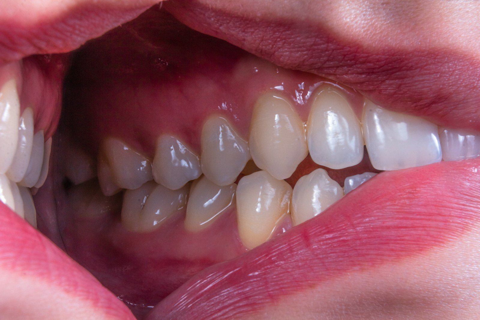
A pale patch has sat on the inside of your cheek for a month. It doesn’t hurt. Your dentist tilts the light, hesitates, and lands on the oldest question in oral medicine: harmless, or the first quiet sign of something that could turn into cancer? A new study asks whether AI oral precancer detection can help answer that faster, and more reliably, than the eye alone.
The 30-Second Version
- A new deep-learning system read clinical mouth photos and told oral precancer (potentially malignant disorders) apart from other lesions better than dentists did on the same images.
- It scored an F1 of 89.9% versus 78.4–82.6% for clinicians, and held up on 1,756 images from other hospitals (F1 ~87%).
- Given to less-experienced dentists as a second opinion, it lifted junior clinicians’ precision by 10.8% — up to the level of their senior colleagues.
- The honest caveat: a photo is not a diagnosis. A biopsy, not a camera, is what confirms precancer — and the data came entirely from academic centers in one country.
It sounds like exactly the early-warning net oral medicine has been missing. But before we hand the mouth-check to an algorithm, it’s worth reading the study closely. In AI oral precancer detection research published in the Journal of Dentistry (2026), Y.Q. Cao and colleagues built a two-stage deep-learning method and tested it against nine clinicians of varying seniority. The guiding question isn’t just “can it score well?” — it’s “does scoring well on photographs mean it can protect a real patient?”
The study, in one glance
The researchers trained a two-stage model on 3,305 clinical intraoral images from one Chinese stomatology hospital. The first stage (they call it CLA OPMD-OOML) decides whether a lesion is an oral potentially malignant disorder or just one of the many other oral mucosal lesions that look alike. The second stage (CLA OPMDs) sorts the precancer into its specific subtypes. They then checked it on 1,756 images from two further centers and ran a blinded, two-step contest: nine dentists — junior, intermediate and senior — diagnosed first alone, then with the AI’s suggestion in hand.
What AI oral precancer detection got right
Two things stand out, and both are real. First, the model didn’t just beat beginners — it out-scored every seniority tier of clinician on the same images, and it did so on two separate axes: flagging which lesions were precancer at all (F1 89.9% vs 78.4–82.6%) and naming the specific subtype (92.2% vs 77.7–80.0%). Second, and more impressive, it mostly kept that performance on 1,756 images drawn from two other hospitals — F1 of 87.3% and 86.9% for the precancer-versus-other task. External validation is the test most dental-AI papers skip, and the one that usually deflates them. This model took it and largely passed.
But can a photograph diagnose precancer?
Here’s where honesty matters. An oral potentially malignant disorder is not cancer; it’s a lesion — a white or red patch, a stubborn ulcer — that carries a raised risk of becoming cancer. And the thing that actually tells a worrying patch from an innocent one isn’t a picture. It’s a biopsy: a sliver of tissue read under a microscope. No camera, however clever, sees the cellular changes that define the diagnosis. What this model does is triage — it decides which patches deserve the knife and the referral, not which ones are malignant.
⚠ “Outperformed clinicians” has an asterisk
The dentists in this study were reading the same flat photographs as the AI — not examining a living mouth, where they could feel a lesion’s texture, ask how long it had been there, or note that the patient chews tobacco. Beating a clinician who has had both hands tied is a narrower win than it first sounds.
An AI that reads the photo is a smoke alarm, not a firefighter: it tells you where to look, but it never puts out the fire or even confirms there is one.
There’s a data limit, too. The model learned and was tested entirely on images from Chinese academic stomatology centers. Oral lesions don’t look identical across the world — pigmentation varies, and the dominant risk habits (betel quid in South Asia, smokeless tobacco elsewhere) produce different lesion patterns. Strong performance in Hangzhou, Chengdu and Shanghai is encouraging; it is not yet proof the tool travels to a clinic in Lagos or Leeds.
Who is accountable when the model is wrong?
Triage cuts both ways. A false “all clear” on a genuinely dangerous patch could delay a cancer diagnosis by months; a false alarm sends an anxious patient for an unnecessary biopsy. The study measured accuracy on a fixed image set, not what happens to real patients downstream when a confident-looking score nudges a busy clinician. The authors are careful to frame the system as decision support — a second reader, not the final word — and that framing is doing important work. The dentist, not the software, still signs the referral.
Does this close the gap, or widen it?
The most quietly exciting result is the collaboration one: junior dentists given the AI’s suggestion improved their precision by 10.8% and intermediate dentists by 5.9%, with the juniors effectively rising to senior-level performance. In a world where most people with a suspicious mouth lesion never see an oral-medicine specialist, a tool that lets a general dentist in an under-served area read a patch like an expert is a genuine equity win — if it reaches those clinics and works on their patients. The risk is the mirror image: if the model is only validated on well-resourced urban populations, it could sharpen care where specialists already cluster and skip the places that need it most.
What this means for you
If you’re a patient
Any mouth patch, lump or ulcer that lasts more than two weeks deserves an in-person look — not a phone photo and an app. AI like this may soon help your dentist decide whether to refer you, but a definitive answer still comes from a specialist and, if needed, a biopsy.
If you’re a clinician
Treat a tool like this as a well-read second opinion that never gets tired, especially for triaging which lesions to refer. Keep examining the patient, not just the image — and remember the model was validated on populations that may not match yours.
The bottom line
This is one of the stronger dental-AI papers of the year: a model that beat clinicians on photographs, survived external validation, and made weaker readers better. But it reads patches, not patients, and it recommends biopsies rather than replacing them. The right destination isn’t an algorithm that diagnoses oral cancer — it’s a sharper, tireless assistant that makes sure the right patches get to the right specialist in time.
Frequently asked questions
Can AI detect oral cancer from a photo of my mouth?
Not by itself. This study’s AI detects oral potentially malignant disorders (precancerous patches) and distinguishes them from other lesions on clinical photographs. It flags which lesions need attention, but a definitive diagnosis still requires an in-person exam and usually a biopsy.
How accurate was the AI compared with dentists?
On the same clinical images, the model reached an F1 score of 89.9% for separating precancer from other lesions, versus 78.4–82.6% for the nine clinicians, and 92.2% versus 77.7–80.0% for naming the specific subtype. It kept F1 scores around 87% on 1,756 images from two other hospitals.
What is an oral potentially malignant disorder?
It’s a lesion — such as leukoplakia (a white patch), erythroplakia (a red patch) or oral lichen planus — that carries an increased risk of turning into oral cancer. Most never do, which is exactly why telling the risky ones apart matters.
Should I use a smartphone app instead of seeing a dentist?
No. The model was tested on standardized clinical images read alongside dentists, not as a consumer self-check. A persistent mouth patch, sore or lump still needs a professional examination.
Is this tool ready to use in clinics now?
Not yet as a standalone. It performed well and even passed external validation, but it was assessed on retrospective images from academic centers in one country. Prospective testing on diverse, real-world patients is the next step before routine clinical use.
Source & author credit
This article interprets, and does not reproduce, the following peer-reviewed study. All figures are the authors’ original findings.
Cao YQ, Zhang JY, Lu M, Shi LJ, Dan HX, Zhu FD, Huang PJ, Zhang GX, Zhang HJ, Chen QM. A deep learning method for diagnosis of oral potentially malignant disorders. Journal of Dentistry. 2026;167:106138. DOI: 10.1016/j.jdent.2025.106138
ORCID — Y.Q. Cao 0000-0003-1822-7118; L.J. Shi 0000-0002-9351-8364; H.X. Dan 0000-0002-9765-0012; F.D. Zhu 0000-0002-9200-9541.
© 2026 Elsevier Ltd (subscription-access journal). Decadentry reports the study’s findings as independent commentary and reproduces no text or figures. Related reading: AI Oral Cancer Screening: Early Warning or False Alarm? · more in Diagnostics & Imaging. Decadentry is an independent educational publication and is not affiliated with the study’s authors.
Leave a Reply