
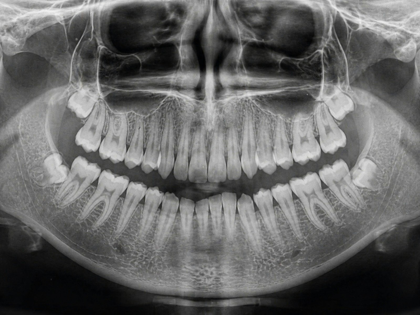
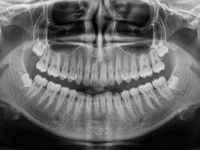
You go in for a routine panoramic X-ray before a new crown. The dentist checks your teeth, your jawbone, maybe a lurking wisdom tooth — and signs off. But low in the corner of that same image, beside the knobbly shadow of your neck vertebrae, sits a faint popcorn-like fleck almost nobody looks for: a carotid calcification, one of the body’s quietest early warnings of a future stroke.
The 30-Second Version
- A 2026 study trained a deep-learning system (YOLOv7) to spot carotid artery calcifications — flecks of hardened plaque linked to stroke and heart disease — on ordinary dental panoramic X-rays.
- The better pipeline flagged 95% of patients who actually had calcifications, versus roughly 21% that general dentists catch with the naked eye.
- But its specificity was only 70%: about 3 in 10 healthy patients were wrongly flagged, often because normal neck anatomy mimics calcium.
- It is one hospital, 580 X-rays, no external validation — a reason to look twice and refer, not a diagnosis.
It sounds like a free bonus from a scan you were getting anyway. But the claim deserves scrutiny. In Imaging Science in Dentistry (2026), Yuta Mitsuya and colleagues at Aichi Gakuin University built two deep-learning detectors — one scanning the whole panoramic image, one zooming first into the neck — and tested them on 580 radiographs. The real question is not just “can AI see the fleck?” but “how often does it cry wolf, and who acts when it does?”
The study, in one glance
The team assembled 580 panoramic radiographs: 290 from patients with confirmed carotid artery calcifications (215 on both sides, 75 on one) and 290 age- and sex-matched controls, collected between 2008 and 2023. Two oral radiologists set the reference standard, and CT scans independently confirmed calcification status in 255 of the cases that made up the validation and test sets. Both detectors used the YOLOv7 network. “System 1” searched the entire image; “System 2” first cropped the bilateral cervical regions where carotid plaques appear, then looked within them.
The promise: spotting carotid calcification a dentist would miss
Carotid artery calcifications matter because they are associated with stroke, cerebral infarction, and coronary heart disease — the plaque you can see is a proxy for the plaque you can’t. The striking part is the human baseline: trained oral radiologists detect these flecks with sensitivity above 0.80, but general dentists, who read most panoramics, catch only about 21%. Against that, System 2 reached 95% patient-level sensitivity, and its side-by-side read of the neck earned an AUC of 0.89 — the strongest result in the paper. In other words, the tool is best at exactly the job humans do worst: not missing the plaque that is there. This is the logic of opportunistic screening — a bonus health signal pulled from an image taken for another reason, much like the AI osteoporosis flags we covered in a 2025 meta-analysis.
How many false alarms is that 95% hiding?
Here is where skepticism earns its keep. System 2’s specificity was just 70%, and its precision (the share of its alerts that were real) hovered around 0.67 — the models placed roughly 30 to 35 bounding boxes on calcium that wasn’t there. The culprits were anatomical look-alikes: the hyoid bone, cervical vertebrae, and the thyrohyoid ligament all cast calcified shadows that resemble a plaque. A 95% catch rate paired with 70% specificity means the system is eager — it rarely misses, but it points at healthy necks often enough that every flag needs a human to confirm it.
⚠ A flag is not a finding
At 70% specificity, a positive result from this system is a prompt to look again, not evidence of disease. Acting on an unconfirmed alert risks worrying patients — and sending them for workups — over the shadow of a neck bone.
It behaves like a smoke alarm that also goes off for burnt toast: you want that sensitivity, but you don’t evacuate the building every time it chirps.
The evidence base is also thin by design. All 580 images came from one institution; not every case was CT-confirmed; the test set was small (80 images, 78 calcifications); and there was no external validation on other hospitals or scanners. The authors are direct about it, naming the limited size and single-center source as central weaknesses. Impressive numbers from one hospital are a hypothesis, not a guarantee of real-world behavior.
Who acts when the dental X-ray raises a medical alarm?
Finding a calcified carotid in a dental office opens an accountability gap technology can’t close. A calcification on a panoramic image is not a diagnosis of carotid stenosis; confirming it needs a medical pathway — a physician referral, typically a neck ultrasound. The detector is only useful if there is an agreed route from “the software highlighted something” to “a doctor evaluated it.” Without that handoff, a flag either gets ignored or spreads alarm with no one responsible for the next step.
Who benefits — and who never gets scanned?
Opportunistic screening has an equity blind spot: it only helps people already getting panoramic X-rays — patients with regular access to dental care. The populations at highest cardiovascular risk often have the least access to a dentist, so a tool that turns routine imaging into stroke-risk screening could quietly widen the gap it appears to narrow, delivering extra warnings to the already-served while missing those who never sit in the chair.
What this means for you
If you’re a patient
You don’t need to request this scan or fear your next X-ray. If a dentist ever mentions a possible calcification near your neck on a panoramic, treat it as a reason to ask your physician about a carotid check — a useful nudge, not a diagnosis.
If you’re a clinician
Tools like this reduce misses but aren’t trustworthy alone. Verify every positive on the image, route it through a referral pathway, and wait for externally validated, multi-center results before leaning on the numbers.
The bottom line
A dental X-ray that quietly screens for stroke risk is a genuinely appealing idea, and AI clearly sees these flecks better than the average dentist. But a 95% catch rate rides on a 70% specificity and a single hospital’s data. This is an assistant that taps the radiologist on the shoulder — not an oracle that delivers verdicts. Used that way, it could save lives; mistaken for certainty, it just manufactures false alarms.
Frequently asked questions
Can a dental X-ray really detect stroke risk?
Not directly. A panoramic X-ray can show carotid artery calcifications — hardened plaque that is statistically associated with stroke and heart disease. It is a risk marker that warrants a medical check, not a prediction or diagnosis of stroke.
How accurate was the AI at finding carotid calcification?
In this 2026 study, the stronger system correctly flagged 95% of patients who had calcifications (sensitivity) but had 70% specificity, meaning roughly 3 in 10 healthy patients were wrongly flagged. Its detection of calcified regions was often confused by normal neck anatomy.
Should I ask my dentist to screen me for this?
No need. The system is a research prototype, not an approved screening tool, and it was tested at only one hospital without external validation. If a calcification is ever noted on your imaging, ask your physician whether a carotid ultrasound is warranted.
Why do false positives happen?
Several normal structures in the neck — the hyoid bone, cervical vertebrae, and the thyrohyoid ligament — can calcify or cast dense shadows that look like arterial plaque, so the AI sometimes marks healthy anatomy as a calcification.
Does this replace a carotid ultrasound or CT?
No. Ultrasound and CT remain the confirmatory tests for carotid disease. The AI’s role, if validated, would be to opportunistically flag suspicious images so patients can be referred for those proper investigations.
Source & author credit
This article interprets, and does not reproduce, the following peer-reviewed study. All figures are the authors’ original findings.
Mitsuya Y, Kuwada C, Yang S, Kise Y, Mori M, Takashi Y, Nishiyama M, Ishikawa N, Naitoh M, Ariji E. Side- and patient-based performance of a deep learning system based on the results of individual detection of carotid artery calcifications on panoramic radiographs. Imaging Science in Dentistry. 2026;56(1):83-92. DOI: 10.5624/isd.20250232
ORCID — Mitsuya 0009-0003-1107-4961 · Kuwada 0000-0002-0065-5192 · Yang 0000-0001-5400-2667 · Kise 0000-0002-5890-2950 · Mori 0000-0003-0275-8707 · Takashi 0009-0007-1144-5506 · Nishiyama 0000-0002-9552-8080 · Ishikawa 0009-0004-0529-616X · Naitoh 0000-0001-8134-7679 · Ariji 0000-0002-5245-5395
Published open access under a Creative Commons Attribution-NonCommercial (CC BY-NC 4.0) licence. Decadentry articles are checked against their primary source per our editorial standards, and sit within our Diagnostics & Imaging research hub. Decadentry is an independent educational publication and is not affiliated with the study’s authors.
Leave a Reply