

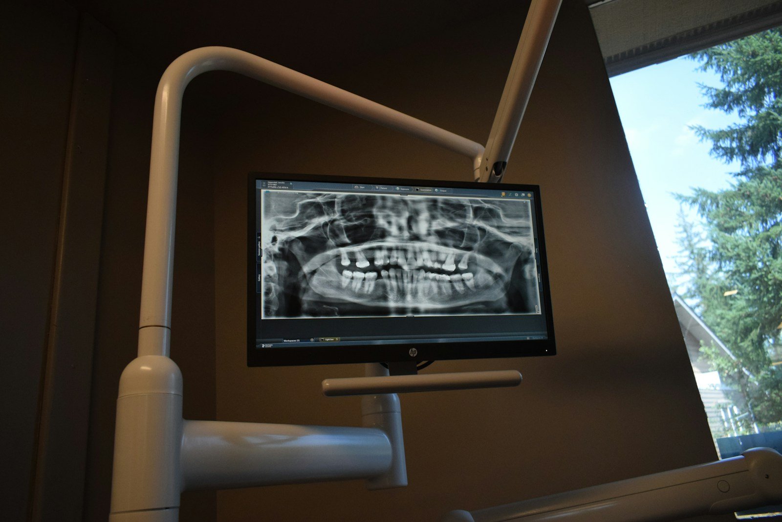
Somewhere in a dental school’s archive sit tens of thousands of panoramic X-rays, each a faint grey map of jawbone and roots. To teach a computer to spot the little dark shadows that betray a dying tooth, a specialist first has to sit down and trace every one of those shadows by hand, thousands of times over. It is the most tedious, expensive bottleneck in dental imaging AI. A new experiment in AI periapical lesion detection asks a cheeky question: what if we just… didn’t?
The 30-Second Version
- A Brazilian team trained deep-learning models to flag periapical bone rarefactions — the dark patches at a root tip that signal an infected or dying tooth — on panoramic X-rays, with nobody hand-drawing a single lesion outline.
- Fed only whole-image “yes/no” labels across 2,527 radiographs, the best model (a fine-tuned Xception network) reached an F1-score of 64.7%.
- That is a long way from the 90%-plus figures headline AI studies love to quote — and the authors are refreshingly upfront that this is a feasibility study, not a finished product.
- The honest caveat: it works as a proof of concept, but it ran on one institution’s images, for a simplified two-way call, and still needs outside validation before it goes anywhere near a clinic.
Skipping the annotations sounds like a corner worth cutting. But cutting it has a price. The study — da Silva-Filho and colleagues, published in Oral Surgery, Oral Medicine, Oral Pathology and Oral Radiology in 2026 — set out to measure exactly how much accuracy you sacrifice when you spare your experts the annotation grind. Their guiding question: can a model learn to see periapical disease from cheap labels alone, and if so, how well?
The study, in one glance
The researchers assembled a retrospective set of 2,527 panoramic radiographs and had experts classify each one, by consensus, using internationally accepted radiographic criteria — but only at the whole-image level, with no boxes or outlines drawn around the lesions themselves. They then pitted several pre-trained architectures against each other, both convolutional neural networks (CNNs) and the newer vision transformers, with fine-tuning and attention mechanisms, and scored them on accuracy, recall, specificity and F1-score. Because some categories were too rare to learn from, the analysis was narrowed to the two most common classes — a binary, present-or-absent call.
The quiet appeal of a lazy label
Here is the part worth getting excited about. Annotation is the hidden tax on almost every medical-imaging AI: before a model can learn, humans have to mark up the data, and marking up a periapical lesion means a trained eye painstakingly outlining a faint smudge of thinned bone on image after image. Image-level labels — just “this film shows disease” or “it doesn’t” — are an order of magnitude cheaper to produce. Showing that a model can still learn something useful from them lowers the barrier to building dental datasets in the first place. The study also delivers a tidy piece of engineering news: the older, unfashionable CNN (a fine-tuned Xception) beat the trendier vision transformers, and did it with low variability between training runs. In a field addicted to the newest architecture, “the boring one won, and won consistently” is genuinely useful to know.
But is this AI periapical lesion detection any good?
An F1-score of 64.7% is, bluntly, middling. F1 is the harmonic mean of precision (when the model says “disease,” how often it’s right) and recall (of the real cases, how many it catches). A score in the mid-60s means a meaningful slice of calls land wrong in one direction or the other. For a condition where a miss can mean an undiagnosed infection quietly eating bone, and a false alarm can mean needless worry or even unnecessary retreatment, that margin is not a rounding error.
⚠ What the score doesn’t tell you
Because the model was trained on whole-image labels, it can only say “somewhere on this X-ray there may be a rarefaction” — not where. There is no box, no arrow, no pointer to the offending root. A dentist still has to hunt the film down tooth by tooth. The cheap label buys a flag, not a map.
It’s a smoke alarm that tells you the building is on fire but not which floor — useful, but you’re still searching room to room.
The evidence base is early-stage, and the authors say so plainly. The data came from a single institution and was gathered retrospectively; the task had to be collapsed to two classes because the others were too rare to train on, so the harder, clinically richer job of grading severity wasn’t attempted. And the paper closes on the line every honest imaging study should: external validation — testing on images from other clinics, scanners and populations — is still required before anyone can trust it to generalize. This is the same unglamorous gap that keeps tripping up AI for reading apical lesions on an X-ray.
Who’s accountable when the cheap model is wrong?
A feasibility model is not a diagnosis, and the real risk with a cheap, easy-to-build tool is scope creep — a 64.7% flag quietly being treated as if it were a verdict. The responsible reading is the one the authors themselves take: the system is an interpretable research probe that still hands the final call to a clinician looking at the actual film. The danger isn’t the honest score on the page; it’s the temptation, once a tool is cheap enough, to lean on it harder than its numbers deserve.
Could cheap labels widen access — or just cheap mistakes?
This is where annotation-free methods are genuinely promising. The places that most need help reading radiographs — smaller schools, rural clinics, under-resourced health systems — are exactly the ones that can’t spare specialists to spend months outlining lesions. A method that learns from labels a busy clinic can actually produce could democratize who gets to build local AI at all. But a model that has only ever seen one institution’s images, graded by one labeling scheme, can carry that narrowness everywhere it’s deployed. Cheap to build can also mean cheap to be wrong — at scale, in the communities least able to catch the error. It’s a reminder that lives in Decadentry’s wider coverage of AI reading dental radiographs: a benchmark is not a bedside.
What this means for you
If you’re a patient
Nothing in your dentist’s office changes today. This is lab-stage research, not a product. Whether a tooth needs a root canal is still decided by a dentist reading your X-ray — usually alongside your symptoms and sometimes a second, more detailed image — not by an app scoring the film.
If you’re a clinician
The headline isn’t the 64.7% — it’s that image-level labels got the model two-thirds of the way with none of the annotation burden, and that a workhorse CNN beat the fashionable transformer. If you’re assembling a departmental dataset, that’s a cost lesson worth banking. Treat any such output as a prompt to look harder, never as the answer.
The bottom line
The real finding here isn’t a number — it’s a trade. You can buy a dental AI cheaply, in expert hours saved, but you pay for it in certainty. 64.7% isn’t the sound of failure; it’s the honest exchange rate between effort and accuracy, printed in a journal for once instead of buried under a press release. The model is a research probe, not a second opinion — an assistant that might one day whisper “look again at this film,” never an oracle that declares the tooth dead. And the authors, to their credit, say exactly that.
Frequently asked questions
What is a periapical bone rarefaction?
It’s a dark (radiolucent) patch of thinned bone at the tip of a tooth root on an X-ray. It usually means the tooth’s nerve has died or become infected and the inflammation has begun dissolving the surrounding bone — the classic radiographic sign that a tooth may need a root canal or extraction.
What does “annotation-free” mean, and why does it matter?
Normally, training a lesion-detecting AI means experts hand-draw an outline around every lesion in thousands of images — slow and expensive. “Annotation-free” here means the model was trained only on a single whole-image label (does this X-ray show a rarefaction or not?), skipping the drawing entirely. It slashes the labeling cost but gives the model less to learn from and no sense of location.
Is an F1-score of 64.7% good?
It’s middling. F1 balances how often the model is right when it says “yes” (precision) against how many real cases it catches (recall). 64.7% sits well below the high-80s-to-90s that screening tools generally aim for, and far below the 90s that heavily annotated models often report. For a feasibility study it’s a reasonable proof of concept; for a clinical tool it isn’t ready.
Can this AI diagnose my tooth?
No. It’s single-institution lab research that flags an image as possibly containing a rarefaction without saying where, and it still needs external validation. Diagnosis stays with a dentist reading your X-ray, usually alongside your symptoms and sometimes a second, more detailed image.
Why did an older CNN beat a vision transformer here?
Vision transformers are the trendy architecture, but they’re data-hungry. On a mid-sized dataset with only image-level labels, the older convolutional network (a fine-tuned Xception) learned more efficiently and gave steadier results — a reminder that newer isn’t automatically better.
Source & author credit
This article interprets, and does not reproduce, the following peer-reviewed study. All figures are the authors’ original findings.
da Silva-Filho JE, Silva CM, Tacchi G, Rodrigues-Fontenele I, Bôtto-Marques M, de Aguiar AWO, Rebouças Filho PP, de Albuquerque DF, Gurgel-Filho ED. Annotation-free deep learning approach for detection of periapical bone rarefactions on panoramic radiographs: a feasibility study. Oral Surgery, Oral Medicine, Oral Pathology and Oral Radiology. 2026;142(5):846–856. DOI: 10.1016/j.oooo.2026.05.007
ORCID — da Silva-Filho JE: 0000-0002-4146-9134 · Silva CM: 0009-0005-1740-2254 · Tacchi G: 0009-0009-7982-2456 · Rodrigues-Fontenele I: 0009-0008-5288-4318 · Bôtto-Marques M: 0009-0007-0815-9971 · de Aguiar AWO: 0000-0001-5437-0945 · Rebouças Filho PP: 0000-0002-1878-5489 · de Albuquerque DF: 0000-0002-5691-3824 · Gurgel-Filho ED: 0000-0002-5772-446X
Published open access under a Creative Commons Attribution 4.0 (CC BY 4.0) licence. Decadentry’s articles are checked against the primary source under our editorial standards. Decadentry is an independent educational publication and is not affiliated with the study’s authors.
Leave a Reply