

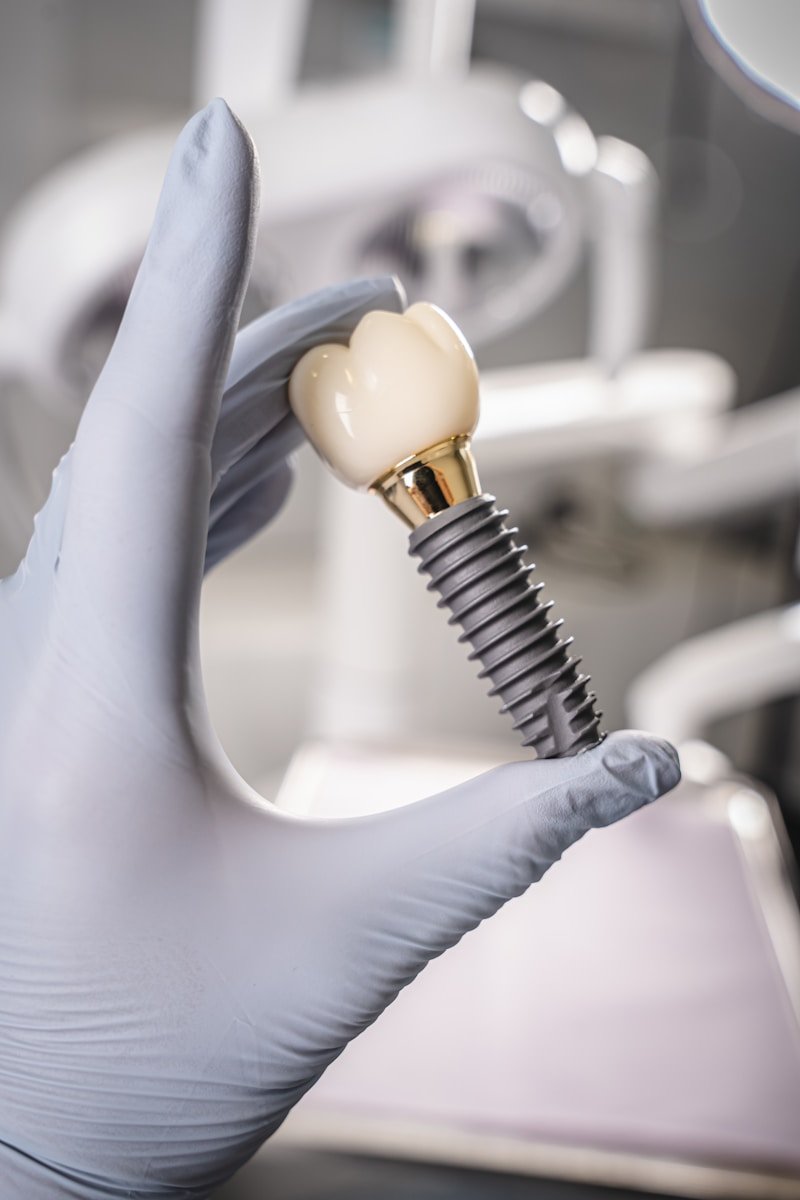
Point a phone at a toothless upper jaw, shoot a short video, and let a neural network rebuild it in three dimensions — accurately enough to seat six implants and screw a full bridge onto them. It sounds like the kind of claim that belongs in a product launch, not a lab. So a team led by the University of Michigan put a version of it on a bench and measured, to the micrometre, exactly how far a smartphone implant scan fell short.
The 30-Second Version
- Researchers turned ordinary smartphone videos of a full-arch implant model into 3D scans using a deep-learning reconstruction model, then measured how true those scans were.
- With custom 3D-printed scan bodies, the smartphone scans strayed about 51 µm from the truth — nearly half the error of off-the-shelf scan bodies (about 81 µm).
- But a conventional impression (~23 µm) and photogrammetry (~27 µm) were still roughly twice as true, and photogrammetry was about six times more precise.
- It is one plastic model on a lab bench — one arch, ten scans per method, no mouth, no saliva. A proof of concept, not a product.
It sounds revolutionary: the scanner that fits in your pocket. But the number that matters in full-arch implant work is measured in microns, and that is where enthusiasm meets physics. In a new study in Clinical Oral Implants Research (2026), Yuqing Lu and colleagues at the University of Michigan, ACTA Amsterdam and the University of Zurich asked a narrower, more honest question than “can a phone scan a mouth?” They asked: if you are going to try, does the marker you point the phone at — the scan body screwed onto each implant — decide whether the whole idea works?
The study, in one glance
The team took a single maxillary edentulous model fitted with six implant analogues and captured it four ways, ten times each: a conventional splinted open-tray impression (the long-standing reference), photogrammetry with a dedicated Icam4D system, and two smartphone-video approaches reconstructed into 3D by a deep-learning model (Neuralangelo) — one using stock PEEK scan bodies, the other using custom scan bodies that were 3D-printed with distinct geometric features and spray-coated to help the algorithm. Then they measured trueness (how close to reality) and precision (how repeatable) as root-mean-square, linear and angular deviations.
Why a smartphone implant scan is even in the conversation
A full-arch intraoral scanner or a photogrammetry rig is a five-figure instrument, and the skill to use it well is not evenly distributed. A smartphone is already in almost every pocket on earth. So the appeal is obvious: if a $0 camera plus a clever algorithm could capture six implant positions accurately enough, digital implant dentistry would suddenly reach clinics — and countries — that cannot justify the hardware. The genuinely interesting finding here is not that the phone worked; it is what made it work better. Simply swapping the generic PEEK scan bodies for custom ones — 3D-printed with sharper geometric features and a matte coating the algorithm could latch onto — nearly halved the trueness error, from about 81 µm to 51 µm, and tightened precision by a similar margin, with every comparison statistically significant. In other words, the camera was not the bottleneck. The thing you point it at was.
But is 51 microns good enough for a full arch?
Here is the uncomfortable part. Full-arch implant restorations are brutally unforgiving of error. When a single rigid bridge is screwed onto four, six or eight implants, every implant has to sit where the scan said it would; a framework that does not fit passively loads the bone and the screws unevenly, and that misfit is implicated in screw loosening, component fracture and, over years, bone loss. Against that standard, the smartphone’s best result — 51 µm with custom scan bodies — was still roughly twice the error of a conventional impression (23.1 µm) and photogrammetry (27.1 µm). On precision, the gap was starker: photogrammetry repeated itself to within about 7.9 µm and 0.07°, while the smartphone method hovered near 50 µm. For the single most demanding job in implant prosthetics, “about twice the error of the reference” is not a rounding detail.
⚠ One model is not one mouth
This was an in-vitro study: a single 3D-printed maxillary model with six implant analogues, scanned on a bench. There was no soft tissue, no saliva, no tongue, no limited mouth opening, and no patient movement — all of which make real intraoral scanning harder, not easier. The lab numbers are a ceiling, not a clinical promise.
A phone-camera panorama is dazzling for a holiday photo and useless for pouring a building’s foundation. The question was never whether the image looks good — it is whether it holds its measurements.
The validation limits matter as much as the headline. One model means we cannot know how the method behaves across different arch shapes, implant angulations or numbers of implants. Ten scans per group is enough to show a difference between scan bodies but thin for pinning down real-world reliability. And the deep-learning reconstruction itself — a research model, not a cleared medical device — needs its own scrutiny before anyone trusts it with a patient’s bone.
Who is accountable if the bridge does not fit?
Digital workflows quietly spread responsibility across a chain — the camera, the reconstruction model, the scan-body design, the lab that mills the framework. When a conventional impression fails, the error is usually visible and the chain is short. When a learned model silently interpolates a surface it could not see cleanly, the failure can be invisible until the finished bridge is tried in and does not seat. That is exactly why the authors frame this as early-stage: a method that looks promising on a bench still has to prove it fails safely, loudly and predictably before it belongs anywhere near a surgical case. It is a close cousin of the trade-offs we flagged when deep learning drafts an implant plan straight from a CBCT scan — impressive on familiar data, shakier the moment conditions change.
Could this widen access — or just add noise?
The equity case cuts both ways. If a refined version of this approach ever reached, say, 25–30 µm reliably, a smartphone-based capture could bring digital implant records to clinics that will never own a photogrammetry system — a real democratising win. But a tool that is “good enough to look convincing” and “not quite good enough to fit” is arguably worse than no tool at all, because it invites confident errors in exactly the settings with the least backup. Access only helps if the thing being accessed is accurate. This study’s most useful contribution may be the reminder that getting there is an engineering problem about markers and reconstruction, not a marketing problem about megapixels.
What this means for you
If you’re a patient
No one is scanning your implants with a phone yet, and this study is a reason to be glad about that. For a full-arch case, a conventional impression or photogrammetry remains the accurate choice. If a clinic ever offers a “smartphone scan,” ask what it has been validated against and whether the final bridge fit was checked — accuracy here is measured in microns, not vibes.
If you’re a clinician
The transferable lesson is that scan-body design is a lever, not a given: custom geometry nearly halved the error. For now this stays in the lab, but it is worth watching as a low-cost capture path for low-resource settings — provided any future version is benchmarked against photogrammetry on real patients, across varied arches, before it touches a framework.
The bottom line
A smartphone plus a neural network can now rebuild a full implant arch in 3D, and clever scan-body design nearly halves its error — a genuinely clever result. But “half as wrong as it used to be” is still about twice as wrong as the tools we already trust. For now the phone is an eager apprentice with real promise, not a replacement for the instruments that keep a full-arch bridge seated for a decade.
Frequently asked questions
Can a smartphone really scan dental implants accurately?
In this lab study it could rebuild a full-arch implant model in 3D to within about 51 µm of the truth using custom scan bodies — impressive, but still roughly twice the error of a conventional impression (23 µm) or photogrammetry (27 µm). It was a single plastic model scanned on a bench, not a patient’s mouth, so it is an early proof of concept rather than a clinic-ready method.
What is a scan body, and why did it matter so much?
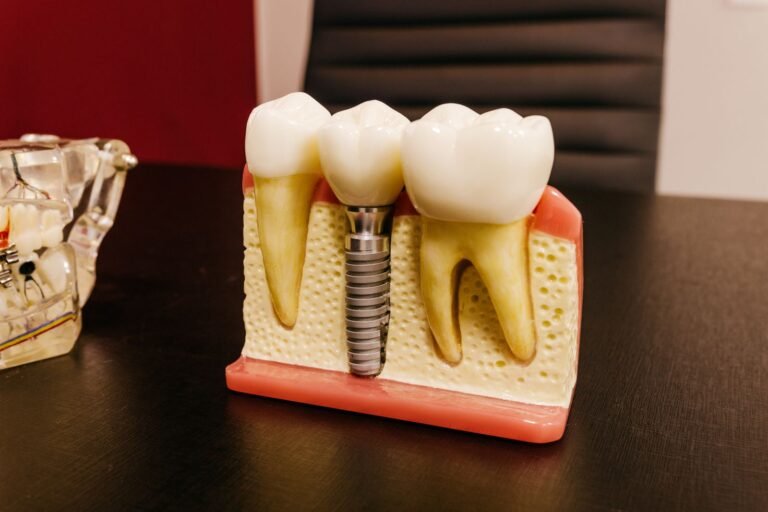
A scan body is a marker screwed onto each implant so a scanner or camera can locate it precisely. The study found that replacing generic PEEK scan bodies with custom 3D-printed ones — sharper geometric features, a matte coating — nearly halved the smartphone method’s error (from ~81 µm to ~51 µm). The camera was not the limiting factor; the marker it photographed was.
Is a smartphone scan as good as a conventional impression?
Not yet. For full-arch implant work the smartphone method was about twice as inaccurate as a conventional splinted impression and far less precise than photogrammetry (which repeated to within ~8 µm). Full-arch bridges are extremely sensitive to tiny errors, so that gap is clinically meaningful.
Could phone-based scanning improve access to implant care?
Potentially — a smartphone costs nothing extra next to a five-figure scanner, so a reliable version could reach clinics that cannot afford dedicated hardware. But that only helps if accuracy improves; a scan that looks convincing yet does not fit could do more harm than good in settings with the least backup.
When might this reach real dental practices?
No firm timeline. The authors describe it as early-stage and in vitro. Before it could be used on patients it would need validation on real mouths across many arch shapes and implant configurations, a cleared reconstruction model, and proof that the final restorations fit — not just that the scans look right.
Source & author credit
This article interprets, and does not reproduce, the following peer-reviewed study. All figures are the authors’ original findings.
Lu Y, Yu J, Liu F, Joda T, Li J. Scan Body Optimisation for Enhancing Full-Arch Implant Scanning Accuracy Using Smartphone Video and a Deep Learning Model: An In Vitro Study. Clinical Oral Implants Research. 2026; advance online publication, e70201. DOI: 10.1111/clr.70201
ORCID — Yuqing Lu (0000-0003-2312-720X) · Jiayi Yu (0000-0001-6455-3366) · Fei Liu (0000-0002-2646-3009) · Tim Joda (0000-0002-1338-5419) · Junying Li (0000-0002-5782-2051)
Published under a Creative Commons Attribution (CC BY 4.0) licence. Figures above are Decadentry’s own interpretation of reported data, not reproductions. Decadentry is an independent educational publication and is not affiliated with the study’s authors. Verified against the study’s published abstract and metadata (full text not yet available via PMC). See our editorial standards and more implants & oral surgery explainers.
Leave a Reply