

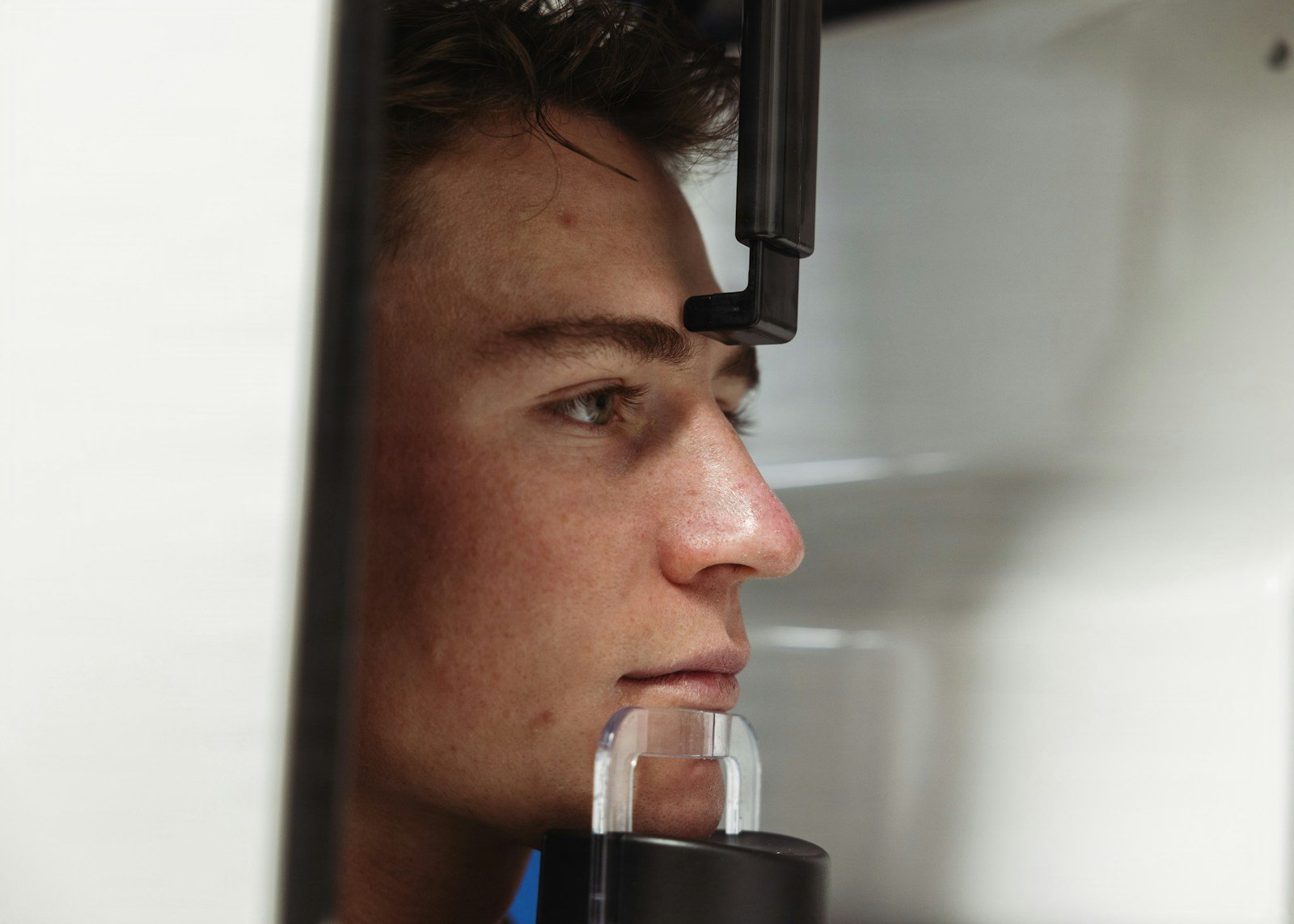
Before a surgeon can graft bone into your upper jaw for a dental implant, someone has to study a paper-thin sheet of tissue lining your maxillary sinus and measure how swollen it is. Too thick, and the operation gets riskier. A new system built for AI maxillary sinus detection says it can read that membrane from a 3D cone-beam CT scan and sort its thickness almost perfectly — 99.43% of the time. That is the kind of number that should make you lean in, and then squint.
The 30-Second Version
- A hybrid deep-learning model sorted maxillary sinus membrane thickening on CBCT scans into four bands (healthy, 2–5, 5–10, and over 10 mm) with 99.43% accuracy.
- It bolted three “attention” modules onto a DenseNet121 network; across five data splits it scored between 98.86% and a perfect 100%.
- Why it matters: a swollen sinus membrane raises the risk of tearing it — or triggering sinusitis — during the “sinus lift” surgery many upper-jaw implants require.
- The catch: it learned from 874 images, one scanner, one clinic, with no outside validation — and a near-perfect score on a small, tidy dataset is a flag, not a finish line.
It sounds like a solved problem. But the claim comes from one specific experiment, and the details decide what it means. Writing in the Journal of Clinical Medicine, Furkan Talo and colleagues at Fırat University in Turkey built a model to detect and classify maxillary sinus membrane thickening on cone-beam CT (CBCT) images — the same kind of 3D scan an AI might one day use to draft the implant plan itself. They ran a DenseNet121 convolutional network through three attention mechanisms in parallel, combined the results, and reported 99.43% accuracy. The question is not whether that number is real. It is what the number does, and does not, tell you.
The study, in one glance
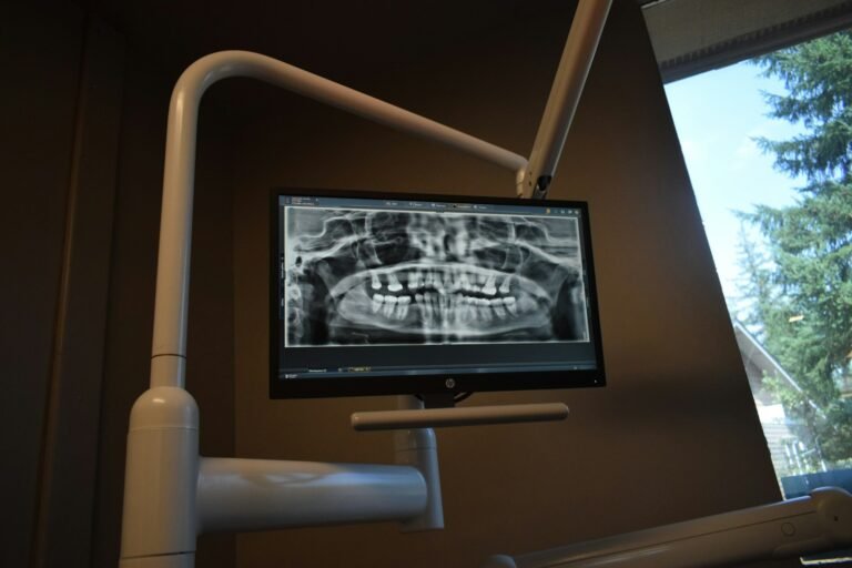
This was a retrospective, single-center study. The team screened 30,000 CBCT scans and built a labeled set of 874 images spread across four thickness classes: 294 healthy (under 2 mm), 181 at 2–5 mm, 190 at 5–10 mm, and 209 over 10 mm. They split the data at the patient level, so no single patient’s images appeared in both the training and testing sets — a genuine mark of care that prevents a model from “cheating.” The architecture used DenseNet121 as a backbone, then passed its features through three attention modules — Squeeze-and-Excitation, CBAM, and Efficient Channel Attention — in parallel before a final classifier. Performance was measured with five-fold cross-validation.
What AI maxillary sinus detection gets right
The clever part earned its keep. Adding the three attention modules nudged DenseNet121’s own 98.86% up to 99.43%, and both comfortably outscored EfficientNetB0 and MobileNetV3-Large (97.14% each) and ResNet50 (91.43%). The model was fast, too — about 4 milliseconds per image — and the patient-level split and cross-validation are the right habits. The clinical target is real and specific: in earlier work, a membrane thicker than 5 mm blocked the sinus’s natural drainage opening in 59.3% of patients, raising the risk of sinusitis after surgery. A healthy membrane is only about 0.8 to 1 mm thick, and measuring it by hand, slice by slice, is slow and varies with the reader’s experience. A consistent, instant first read is a worthy goal.
Does 99.43% mean it’s ready for your jaw?
Not yet — and the reason is in the fine print. That near-perfect accuracy came from 874 images produced on a single CBCT machine (a Planmeca ProMax 3D Mid) at one university clinic. When a model scores a flat 100% on one of its data folds, as this one did, that is often a sign the dataset is clean and neatly separable, not that the real world has been conquered. The authors say so plainly: single-center data, no external validation, prospective clinical testing still to come.
⚠ A number without a second clinic
A model that aces its own clinic’s scans can stumble on a different scanner, a different resolution, or a different patient population. Until it is tested on outside data, 99.43% describes this dataset — not your dentist’s.
It’s like a student who aced every practice test — written by their own teacher. Impressive, until they sit an exam someone else set.
There is a second, subtler limit. The model sorts thickness into four preset bands from pre-selected 2D cross-sections. It is not hunting for the sinus in the first place, measuring the single thickest point across the whole 3D volume, or deciding what to do about what it finds. Real membranes do not respect the 2, 5, and 10 mm lines: a 4.9 mm membrane and a 5.1 mm membrane sit in different boxes but look almost identical, and that boundary is exactly where a classifier is most likely to slip.
Who owns the call when the scan is borderline?
The model outputs a label, not a judgment. A borderline membrane — scarred by old infection, carrying a harmless retention cyst, or stiffened by fibrosis — still needs a clinician to read it in context. The study itself notes that the membrane’s elasticity and fibrosis also drive the risk of perforation, and those are things a thickness classifier simply cannot see. Accountability for the surgical plan stays with the surgeon; the AI is a fast first pass, and a confident wrong label is more dangerous than an honest “I don’t know.”
Will it work outside the clinic that built it?
That is the open question. Every image came from one clinic, one machine, one set of acquisition settings, one population. Clinics running older scanners, different voxel sizes, or serving different patients are precisely where an unvalidated model is most likely to drift — and those are often the same clinics with the least access to a specialist for a second opinion. Generalization, not raw accuracy, is the unfinished work here, and it is the work that decides whether a lab result ever becomes a tool you benefit from.
What this means for you
If you’re a patient
If you’re facing an upper-jaw implant and a sinus lift, your CBCT scan and your surgeon’s reading of it still run the show. A tool like this might one day give your dentist a faster, steadier first measurement of your sinus membrane — but it will not replace the clinical judgment that decides whether, and how, to operate.
If you’re a clinician
Promising architecture, honest limitations. Read the 99.43% as an internal-dataset ceiling, not a deployment spec. Before trusting any sinus-membrane classifier, ask for external, multi-center validation, its performance right at the fuzzy 5 mm boundary, and whether it measures true 3D maximum thickness or merely sorts slices you hand it.
The bottom line
A near-perfect score is the beginning of scrutiny, not the end of it. This model is a capable assistant that can flag a thickened sinus membrane quickly and consistently — but it is an assistant that has only ever worked in one building. Trust it the way you’d trust a sharp new resident: useful from day one, supervised until proven elsewhere.
Frequently asked questions
What is maxillary sinus membrane thickening, and why does it matter for implants?
The maxillary sinus — the air space above your back upper teeth — is lined by a thin membrane, normally about 0.8 to 1 mm thick. Infections, allergies, or dental problems can thicken it many times over. Because many upper-jaw implants need a “sinus lift” to add bone, a swollen membrane raises the risk of tearing it or triggering sinusitis, so surgeons check it on a CBCT scan before operating.
How accurate was the AI, really?
On its own test data, it classified membrane thickness into four bands with 99.43% accuracy, edging out a DenseNet121 model at 98.86% and sitting well above ResNet50 at 91.43%. Across five cross-validation folds it scored from 98.86% to a perfect 100%. But all of that was measured on 874 images from a single clinic and a single scanner, with no outside validation.
Can this replace my dentist reading the scan?
No. It sorts thickness into preset categories from slices it is given; it does not locate the sinus, measure the true 3D thickest point, or decide on treatment. The authors describe it as a pre-operative planning aid and say prospective, multi-center testing is still needed before clinical use.
Why be skeptical of a 99% score?
Because near-perfect accuracy on a small, single-center dataset often reflects how clean and separable that particular data is, not how the model will behave on a different scanner or population. Real clinical performance is proven by external validation on outside data — which this study has not done yet.
Source & author credit
This article interprets, and does not reproduce, the following peer-reviewed study. All figures are the authors’ original findings.
Talo F, Duger N, Dagtekin B, Karaduman M, Yildirim M, Talo Yildirim T. Automatic Detection of Sinus Membrane Thickness Using a Hybrid Deep Learning Model Integrating Multiple Attention Mechanisms. Journal of Clinical Medicine. 2026;15(18):6988. DOI: 10.3390/jcm15186988
ORCID — Furkan Talo 0000-0003-4891-7873; Nurullah Duger 0000-0002-0625-3864; Burak Dagtekin 0009-0001-1982-8225; Mucahit Karaduman 0000-0002-8087-4044; Muhammed Yildirim 0000-0003-1866-4721.
Published open access under a CC BY 4.0 license. Study selection and fact-checking follow our editorial standards; see more explainers in Diagnostics & Imaging. Decadentry is an independent educational publication and is not affiliated with the study’s authors.
Leave a Reply