

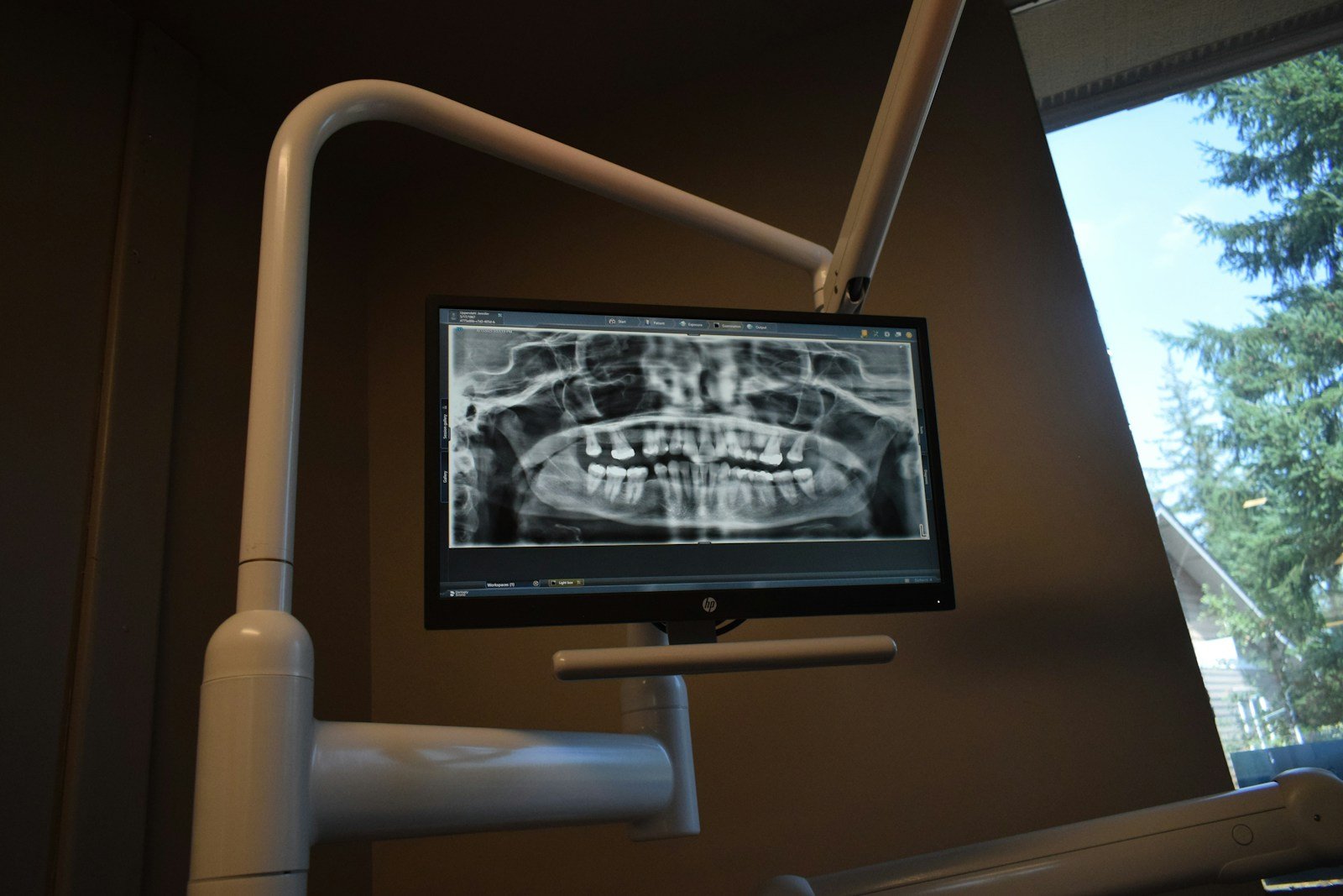
A teenager face-plants off a skateboard and lands in your chair an hour later: split lip, a front tooth that wobbles when she talks. The radiograph goes up on the monitor, and the real question is whether a crack is creeping down into the root — the kind of call that decides between a splint and a lost tooth. Now imagine an AI scanning that same image first. This is exactly the promise of AI dental trauma detection, and a new study put it to the test.
The 30-Second Version
- Researchers trained a modern object-detection model (YOLO26x) to find and label dental injuries on mostly cone-beam CT images, plus some periapical X-rays.
- It was genuinely sharp on the dramatic fractures — about 90% average precision for complicated crown-root fractures and 88% for crown fractures.
- But it badly missed the subtle ones: only 27% average precision for alveolar (jaw-bone socket) fractures and 49% for one crown-root subtype.
- The honest caveat: a single center, internal testing only, and no patient-level separation — so these are promising lab numbers, not a clinic-ready tool.
It sounds like the perfect co-pilot for a chaotic trauma visit. But in a study published in the Journal of Dentistry (2026), Wangyue Dai and colleagues at Xi’an Jiaotong University trained a YOLO26x deep-learning model on 1,256 annotated trauma instances — 1,065 drawn from sagittal cone-beam CT (CBCT) slices and 191 from periapical radiographs — to do something harder than most dental AI attempts: not just “is there damage?” but “what kind of fracture is this, and exactly where?” That fine-grained question is where the interesting cracks appear.
The study, in one glance
This was a retrospective, single-center study designed to develop and internally evaluate the model. The team taught it to localize and classify five trauma subtypes, then measured performance with average precision (AP) — a combined score of how often the model’s boxes are both correct and complete. The results split cleanly into two worlds: the fractures it could see, and the ones it couldn’t.
What AI dental trauma detection gets right
When the damage is obvious, the model is impressive. Complicated crown-root fractures — the painful, visible ones where the break crosses from the crown into the root and exposes the pulp — scored 90.4% average precision. Simple crown fractures hit 88.2%, and root fractures, which are notoriously easy to overlook on a flat 2D X-ray, reached 83.9%. And it did this fast: an average of 3.3 milliseconds per image, which is “faster than you can blink” territory. In a busy emergency room at 2 a.m., a tool that reliably flags the dramatic fractures and highlights exactly where they sit could be a real second pair of eyes — especially for a tired clinician or one who doesn’t see trauma every day.
So why did it miss the quiet injuries?
Because the hardest fractures to see are the hardest for AI to learn. Alveolar fractures — breaks in the bony socket that holds the tooth — scored just 27.3% average precision. One subtype of crown-root fracture landed at 48.9%. Across all five categories, the overall mean average precision was 69.9%, and the stricter mAP@0.5:0.95 score (which demands near-perfect boxes) was only 42.5%, with overall precision of 0.675 and recall of 0.704. Translated: for every ten injuries present, this version would tend to catch about seven and miss three, and roughly a third of its alarms wouldn’t pin the right thing.
⚠ The miss that matters most
Alveolar fractures are both easy to overlook clinically and potentially serious — a missed socket fracture can doom the teeth it supports. An AI that catches the eye-catching breaks but fumbles the camouflaged ones could quietly reinforce the exact blind spot a clinician most needs help with.
A smoke alarm that reliably screams at a kitchen fire but stays silent for a smoldering wire isn’t a safety net — it’s a false sense of one.
Part of the problem is sheer rarity: the model simply had fewer examples of the uncommon subtypes to learn from, and deep learning is only as sharp as the patterns it has seen many times. Part of it is physics — some of these injuries are faint even to a trained human eye, and the CBCT-heavy dataset doesn’t change the fact that an alveolar hairline can hide in the noise.
Can you trust a number built on one dataset?
This is where scientific honesty matters. The study was retrospective and single-center, and — crucially — the authors themselves flag that it was internally evaluated without patient-level separation. That last point is a subtle but important risk: if different images from the same patient end up in both the training and test sets, a model can look smarter than it is, because it has effectively “seen” the test case before. The authors are refreshingly upfront that these are preliminary feasibility findings that “warrant multicenter validation with patient-level separation.” In other words: promising proof of concept, not evidence of clinic-ready performance.
Who does a tool like this actually help?
Trauma doesn’t wait for specialists. A lot of dental injuries first show up in general practices, pediatric clinics, and emergency departments where no oral radiologist is on hand. A reliable detector for the common, dramatic fractures could genuinely raise the floor in exactly those settings. But the equity catch is real: the rare, subtle fractures the model handles worst are also the ones a non-specialist is least likely to catch unaided — so the current tool is strongest where help is least needed and weakest where it’s needed most. Closing that gap, not just publishing a headline accuracy, is the work that counts.
What this means for you
If you’re a patient
After a knock to the mouth, a dentist’s hands-on exam and judgment still decide your care — not a screen. If an AI label appears on your X-ray, treat it as a highlighter, not a diagnosis. And if pain or looseness lingers, ask about a follow-up image: some fractures only reveal themselves days later, and no model fixes that.
If you’re a clinician
A detector like this could triage the obvious and speed up documentation, but its alveolar and subtle-subtype scores say: don’t let a quiet AI lull you. Use it to confirm what you suspect, not to rule out what you don’t — and remember it hasn’t been tested on your scanner, your population, or with clean patient-level splits.
The bottom line
This model is a sharp-eyed assistant for the fractures that already announce themselves, and an unreliable one for the injuries that hide. That’s not a failure — it’s an honest snapshot of where dental trauma AI stands: good enough to help a clinician look, nowhere near good enough to decide. The goal was never an oracle that reads the X-ray for us. It’s a second set of eyes that makes a human’s first set sharper.
Frequently asked questions
Can AI reliably detect a cracked or fractured tooth from an X-ray?
For obvious fractures — crown breaks and complicated crown-root fractures — a modern AI model reached around 88–90% average precision in this study, which is genuinely useful. But for subtle injuries like alveolar (socket) fractures it dropped to about 27%, so it is not reliable across all fracture types yet.
What is average precision, and why not just say “accuracy”?
Average precision (AP) combines how often the AI’s detections are correct with how many true injuries it actually finds, at a set standard for how well its box must overlap the real lesion. It’s a tougher, more honest measure than plain accuracy for “find and label the damage” tasks, because it penalizes both false alarms and missed injuries.
Does this mean AI will diagnose dental trauma instead of a dentist?
No. The study was a single-center, internal proof of concept, and the authors explicitly call for multicenter validation before clinical use. A tool like this is designed to flag and localize possible injuries for a clinician to confirm — it supports the diagnosis, it doesn’t make it.
Why was the AI so much worse at alveolar fractures?
Two reasons: alveolar (jaw-socket) fractures are relatively rare, so the model had fewer examples to learn from, and they are genuinely faint on imaging even for human experts. Less data plus a harder visual target equals weaker performance.
Should I trust an AI reading of my own dental X-ray?
Treat it as a helpful highlight, not a verdict. This kind of model hasn’t been validated on the specific scanner or population you’d encounter, and some fractures only become visible on a repeat image later. Your dentist’s clinical exam remains the deciding factor.
Source & author credit
This article interprets, and does not reproduce, the following peer-reviewed study. All figures are the authors’ original findings.
Dai W, Wu J, Li T, Gong L, Chen X, Lei Y, Li J, Gao S, Lu Z, Cheng B, Chen C. Development and internal evaluation of a deep learning model for fine-grained detection of dental trauma on CBCT-dominant image records. Journal of Dentistry. 2026;176:106984. DOI: 10.1016/j.jdent.2026.106984
ORCID — Lingjuan Gong 0009-0004-8340-287X; Baixiang Cheng 0009-0004-3184-8677; Cheng Chen 0000-0003-3215-9449 (verified via Crossref).
© 2026 Elsevier Ltd. All rights reserved. This is a subscription (non-open-access) article; Decadentry summarizes and interprets it as independent editorial commentary and does not reproduce the original text or figures. Decadentry follows transparent editorial standards and is an independent educational publication, not affiliated with the study’s authors. Related reading: AI Dental Trauma Advice: Grounded Fact or Confident Guess? and the Diagnostics & Imaging research hub.
Leave a Reply