
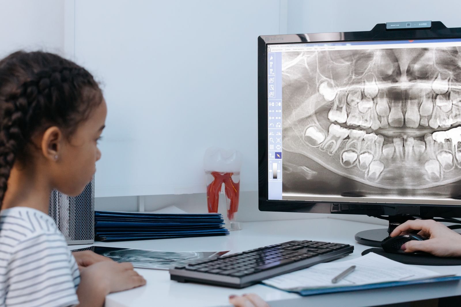
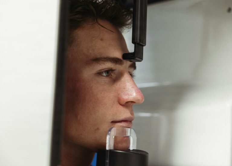
Your child’s orthodontist takes a side-on X-ray of the head to plan braces. Routine, low-dose, over in a second. But that same picture quietly records something no one was looking for — the shape of the airway at the back of the throat, and the jaw that frames it. A new study asks whether an algorithm can read that hidden clue and flag a child who may be choking for air every night.
The 30-Second Version
- Researchers trained a two-stage deep-learning model to flag pediatric obstructive sleep apnea risk from a routine lateral cephalogram — the side-view dental/orthodontic head X-ray.
- On a held-out test set it reached an AUC of 0.945, and with its help dentists’ accuracy rose by up to 23.7 percentage points.
- It is a screening flag, not a diagnosis: a sleep study still confirms the condition.
- The honest caveat: just 188 children from a single hospital, retrospective, with no external multi-center validation yet.
It sounds like a gift — catch a hidden illness from an X-ray you already took. But enthusiasm is not evidence. The study, by Jiayi Zhang and colleagues at the Affiliated Stomatological Hospital of Chongqing Medical University and published in Frontiers in Pediatrics (2026), built an AI sleep apnea screening tool that reads a child’s lateral cephalogram and estimates the odds of obstructive sleep apnea-hypopnea syndrome (OSAHS). The guiding question is not “is this clever?” — it clearly is — but “is it ready to change what happens in a dental chair?”
The study, in one glance
The team retrospectively gathered lateral cephalograms from 188 children (mean age about 10.6 years) seen between January 2021 and October 2025. They split the data into a development set for five-fold cross-validation and an independent hold-out test set of 38 images. The model works in two stages: first an nnU-Net segments the upper airway; then a modified ResNet-34 classifier fuses the whole-X-ray craniofacial view with the cropped airway to decide OSAHS versus not. Children were labelled using the apnea-hypopnea index — in pediatrics, even one event per hour (AHI ≥ 1) counts, a far lower bar than in adults.
The genuinely impressive part
Pediatric OSAHS is common — it affects roughly 1% to 4% of children — and it is easy to miss. Left untreated it is linked to disrupted growth, poor concentration, and behaviour and learning problems. The diagnostic gold standard, overnight polysomnography (a sleep study), is expensive, scarce, and hard to run at scale in children, so many cases never get caught. The appeal here is opportunistic: lateral cephalograms are already taken in orthodontic and dental practice at low radiation dose. If the picture can double as a free first screen, a dentist could spot a high-risk child and refer them for the sleep study they would otherwise never have had. The model’s 0.945 AUC on the test set — well above a whole-image baseline of 0.797 — and a clean airway segmentation (Dice similarity 0.931) suggest it is reading something real: the airway narrowing and jaw features clinicians already associate with childhood apnea, which the authors’ Grad-CAM heatmaps traced back to the adenoid, tonsil, and craniofacial regions.
But how good is 0.945 when only 38 children were tested?
A test set of 38 images is small, and it shows in the confidence interval: the test AUC’s lower bound dips to 0.863, and for some sub-metrics the intervals are far wider. More importantly, the paper is internally inconsistent about its own F1 score — the abstract reports 0.933 while the results table reports 0.824 for the same test set — which is exactly why a careful reader leans on the figure the paper reports consistently (the AUC) and treats the rest with caution. One strong number from one small, retrospective sample is a promising signal, not a settled result.
⚠ “Two hospitals” is really one
The children came from two campuses of a single institution, scanned on the same equipment. That is not external validation. The model has never been tested on a different hospital, a different scanner, or a different population — the step that decides whether a lab result survives contact with the real world.
A cephalogram screen is a smoke alarm, not a fire inspection: useful precisely because it is cheap and everywhere, but it tells you to look closer, not that the house is burning.
There is a subtler problem in how “healthy” was defined. Only 118 of the 188 children had a confirming sleep study; the other 70 controls were cleared by questionnaires, a CBCT scan, and clinical judgement. Mild or silent apnea could easily hide in that control group, teaching the model a slightly blurred version of the truth — a limitation the authors acknowledge.
Does a sharper flag mean a better diagnosis?
The most clinically interesting result was not the AUC but the reader study. Eight dentists read the same 38 X-rays with and without the model’s help; accuracy rose by 16.5 points for juniors and 23.7 points for seniors. That is the right way to frame an AI tool — as something that lifts a human, not replaces one. But the flip side deserves naming: if the model is wrong, it can lift a clinician’s confidence in the wrong direction just as efficiently. Tools that make experts feel more sure are powerful exactly when they are right and dangerous exactly when they are not, and this one has not yet faced a crowd of patients it has never seen.
Who gets the free screen — and who gets missed?
Opportunistic screening only helps the children who already walk through the door with an X-ray. A model like this could extend the reach of a routine image, the same promise behind using dental radiographs to flag stroke risk or bone loss. But it also risks widening a gap: kids who never see an orthodontist — often the same kids with the least access to a sleep clinic — get neither the flag nor the follow-up. A screening tool that rides on dental care inherits dental care’s inequalities unless someone designs around them.
What this means for you
If you’re a patient (or parent)
If your child snores loudly, gasps, or sleeps restlessly, that is worth raising with a doctor regardless of any X-ray. This kind of AI might one day add a prompt from the dental side, but it does not diagnose apnea and is not in your clinic yet. A sleep evaluation remains the real test.
If you’re a clinician
Treat cephalogram-based apnea models as an emerging referral aid, not a diagnostic. The signal is plausible and the reader-study lift is encouraging, but single-center, retrospective performance rarely holds at full strength elsewhere. Watch for prospective, multi-center validation before leaning on it.
The bottom line
This study makes a genuinely appealing case that an X-ray you already have can whisper a warning about a child’s sleep. But a whisper is a reason to listen harder, not a verdict. The model is an assistant pointing at the airway and saying “check this one” — not an oracle declaring the diagnosis. Its job, done well, ends with a referral to a sleep study, not instead of one.
Frequently asked questions
Can an AI diagnose sleep apnea from a dental X-ray?
No. In this study the AI estimated the risk of pediatric obstructive sleep apnea from a lateral cephalogram and reached an AUC of 0.945 on a small test set, but the authors frame it as a screening flag. An overnight sleep study (polysomnography) remains the diagnostic gold standard.
What is a lateral cephalogram?
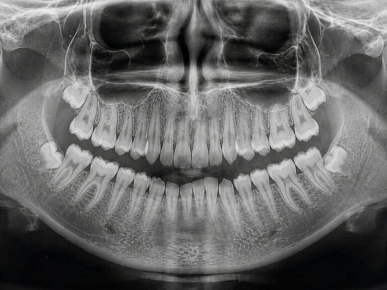
It is a standardized side-view X-ray of the head routinely taken in orthodontic and dental practice to assess jaw and facial structures. Because it also captures the soft tissue of the upper airway at low radiation dose, researchers are testing whether it can double as an apnea screen.
How accurate was the AI sleep apnea screening model?
On its independent 38-image test set the fusion model scored an AUC of 0.945 (95% CI 0.863–0.994), above a whole-image baseline of 0.797. With the model’s help, dentists’ accuracy rose by 16.5 points (juniors) and 23.7 points (seniors). The test set was small and from a single institution.
Why is external validation such a big deal here?
The 188 children came from two campuses of one hospital, scanned on the same machine. AI models often perform worse on images from a different clinic, scanner, or population. Until the model is tested prospectively across multiple centers, its strong score is a promising signal rather than proof of clinical readiness.
Source & author credit
This article interprets, and does not reproduce, the following peer-reviewed study. All figures are the authors’ original findings.
Zhang J, Tan J, Tong X, Wang H, Zhao Y, Song J, Liu Y. Interpretable two-stage deep learning for pediatric obstructive sleep apnea diagnosis using lateral cephalograms. Frontiers in Pediatrics. 2026;14:1817094. DOI: 10.3389/fped.2026.1817094
ORCID — no author ORCID iDs were listed in the article’s Crossref record, so none are cited here.
Published open access under a Creative Commons Attribution 4.0 (CC BY 4.0) license; figures above are Decadentry’s own interpretation, not reproductions. Study reviewed against its primary source per our editorial standards. Filed under Diagnostics & Imaging. Decadentry is an independent educational publication and is not affiliated with the study’s authors.
Leave a Reply