
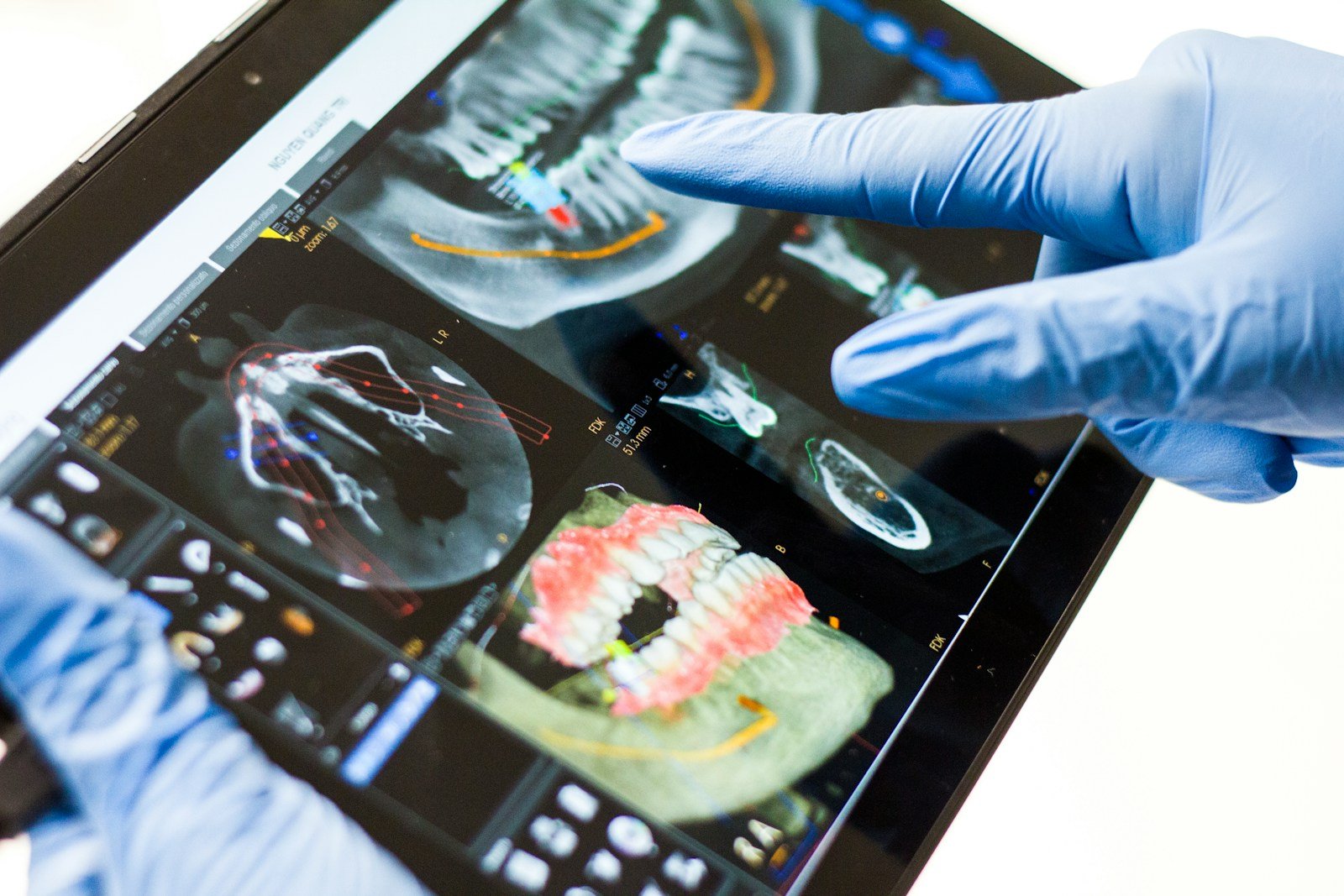
Your implant looks fine. The gum hugging it is maybe a shade pinker than it was at your last cleaning — or maybe that’s the lighting, or your imagination. “A little red” is one of the softest judgment calls in dentistry, and it happens to be the exact signal that separates a healthy implant from one quietly sliding toward peri-implant disease. A team at Shanghai Jiao Tong University decided that guess was too important to leave to tired eyes and variable lighting, so they built a tool for AI peri-implant monitoring that turns the colour of your gum into a hard number.
The 30-Second Version
- Researchers trained AI to read peri-implant gum redness from a routine 3D intraoral scan, producing an objective “Digital Erythema Score” (DES) instead of a dentist’s eyeball guess.
- It flagged inflamed gums well — area-under-the-curve 0.92 in its home clinic and 0.90 in an outside one — and a high score meant a site was over five times more likely to bleed on probing.
- But its ability to pinpoint where the inflammation sat fell sharply outside the home clinic (localization mAP 0.90 → 0.68), and the entire external test was just 23 patients.
- Honest caveat: it was judged against dentists’ visual scores (not biopsy), captured with a professional scanner (not your phone), at a single Chinese centre — so the home-monitoring dream is still a dream.
It sounds like the end of “looks a little inflamed to me.” But the study — Wu and colleagues, published in Clinical Oral Implants Research in November 2026 — is careful to call itself “preliminary,” and that word is doing real work. The guiding question isn’t “can AI see redness?” It’s “can an objective redness score survive leaving the clinic that built it?” Here’s where it holds, and where it slips.
The study, in one glance
The researchers assembled intraoral scans from 200 patients (265 implants) and had two independent experts agree on which peri-implant tissues were inflamed, using the modified Gingival Index (mGI 0 = healthy, mGI ≥ 1 = erythema). A U-Net neural network learned to trace the thin collar of gum right around each implant; a colour algorithm converted that strip into the Digital Erythema Score; and a YOLO object detector tried to draw a box around the reddest, most inflamed regions. They then tested the whole pipeline on a separate, independent set of 23 patients (30 implants) and ran a multi-observer study to see whether the score helped real clinicians judge more consistently.
What AI peri-implant monitoring gets right
The headline strength is consistency. The segmentation was solid (Dice similarity 0.69) and the colour maths was almost perfectly repeatable (intraclass correlation above 0.95) — meaning the score barely changes if you re-run it, which is precisely where human eyes fail. More importantly, the score tracked real biology: a higher Digital Erythema Score multiplied the odds that a site would bleed on probing — the clinical tell of inflammation — more than fivefold (odds ratio 5.32, p < 0.001). And it kept its detection sharpness when it left home: an AUC of 0.92 internally and 0.90 on the outside clinic’s patients is a genuinely good result, not a lab mirage. In the observer study, handing clinicians the DES made dentists of different experience levels agree with each other more — turning a subjective squint into a shared, checkable number.
But does it hold up once it leaves home?
Partly. Detecting that something is inflamed held up; knowing where did not. The YOLO localizer scored a strong mAP of 0.90 on the home clinic’s images and then dropped to 0.68 on the outside clinic’s — nearly a quarter of its precision gone the moment it met unfamiliar scans. That gap is the oldest story in medical AI: a model quietly memorises the look of one clinic’s equipment and lighting, then stumbles on everyone else’s.
⚠ A 23-patient window
The external validation that is supposed to prove a model works in the wild was 23 patients and 30 implants. That is enough to be encouraging and nowhere near enough to be convincing — the confidence interval on that external detection AUC runs from 0.81 to 0.98, a range wide enough to drive a scanner through.
Think of it as a blood-pressure cuff for the gum around an implant: it puts an honest number on a warning sign, but it never tells you the diagnosis.
And the “ground truth” here was two experts’ visual grading, not a biopsy or a bone measurement. So the AI learned to match expert eyes, inheriting whatever those eyes miss. A tool that agrees with dentists is useful; a tool that is right is something you have to prove against harder evidence than another clinician’s opinion.
Does a red flag actually mean disease?
Redness is a symptom, not a verdict. Peri-implant mucositis (reversible gum inflammation) and peri-implantitis (inflammation plus bone loss that can cost you the implant) are different animals, and bone loss doesn’t show up as colour on a surface scan. The DES is an early, visible warning — the smoke, not the fire. That is valuable, because catching mucositis early is the best chance to stop it before bone is involved. But a score that says “inflamed” still needs a clinician with a probe, and often an X-ray, to say what that inflammation means. A separate line of research is trying to predict peri-implantitis before it starts from a baseline X-ray — and it, too, lands as a signal to watch rather than a verdict.
Whose gums was it trained to read?
This is where a colour-based tool deserves extra scrutiny. The whole system was built at one Chinese centre on that population’s tissues, and erythema is, by definition, a judgment about colour against a baseline. Natural gum pigmentation varies widely between people, and a redness score calibrated on one range of tissue tones can misjudge others — over-flagging darker, more pigmented gums or missing subtle inflammation against them. Until a tool like this is validated across skin and mucosal tones, “objective” is a promise, not a guarantee, and the people most likely to be mis-scored are the ones already least represented in the training data.
What this means for you
If you’re a patient
This is a glimpse of future check-ups, not something in your bathroom yet. The scans here came from a professional intraoral scanner, not a phone, and the authors are explicit that home use waits on “validation with patient-accessible imaging devices.” For now the old advice stands: clean around your implants daily and keep your implant maintenance visits — bleeding gums, not an app, are still your earliest honest signal.
If you’re a clinician
A repeatable erythema score is a real addition to implant maintenance — useful for tracking one patient’s tissue over time and for levelling out the disagreement between a new hire and a veteran. Treat it as a documentation and triage aid that sharpens your own exam, not a replacement for probing, radiographs and judgment — and stay sceptical of its localization until it’s proven on your own equipment.
The bottom line
The most grounded thing about this study is its own title: “preliminary.” It shows, convincingly, that an AI can convert the fuzzy clinical phrase “a little red” into a number that moves with real inflammation — a genuine step toward objective, trackable implant care. But converting a sign into a score is not the same as making a diagnosis, and proving it on 23 patients in one clinic is not the same as proving it in the world. The right posture is the one the researchers themselves take: a promising assistant that quantifies what the clinician already looks for — never an oracle that decides.
Frequently asked questions
What is a Digital Erythema Score (DES)?
It’s a number an AI calculates from the colour of the gum tissue surrounding a dental implant on a 3D intraoral scan. A higher score means more redness (erythema), an early sign of inflammation. The goal is to replace a dentist’s subjective “looks a bit red” with a consistent, repeatable measurement.
Can I use AI peri-implant monitoring at home with my phone?
Not yet. In this study the scans came from a professional intraoral scanner, and the authors say home use depends on future validation with patient-accessible cameras. Today it’s a clinic research tool, not a consumer app.
Does a red flag from the AI mean I have peri-implantitis?
No. Redness signals inflammation, which may be reversible mucositis rather than peri-implantitis — the more serious form that involves bone loss. Bone loss doesn’t show up as surface colour, so a high score is a prompt to get a clinical exam and possibly an X-ray, not a diagnosis.
Is the AI better than my dentist’s eye?
It’s more consistent, not necessarily more correct. It barely changes between readings and helped clinicians agree more with each other, but it was trained to match expert visual scores rather than biopsy-confirmed truth, and its ability to pinpoint inflammation dropped sharply at an outside clinic.
Source & author credit
This article interprets, and does not reproduce, the following peer-reviewed study. All figures are the authors’ original findings.
Wu X, Wei X, Lv X, Fu X, He Z, Yin W, Lai H, Shi J. AI Enabled Automated Digital Image Analysis for Peri-Implant Health Monitoring: A Preliminary Diagnostic Study. Clinical Oral Implants Research. 2026; advance online publication (Article e70200). DOI: 10.1111/clr.70200. Prospectively registered: ClinicalTrials.gov NCT07349095.
ORCID — Xinyu Wu 0000-0003-1310-1452; Xindi Wei 0000-0002-0313-339X; Xiaolei Lv 0009-0002-7067-7044; Xiaojiao Fu 0000-0002-9934-5388; Hongchang Lai 0000-0002-6922-2918; Junyu Shi 0000-0002-8241-2572.
© 2026 The Authors. Clinical Oral Implants Research published by John Wiley & Sons Ltd. This is a copyrighted article; Decadentry summarises and does not reproduce it. Every Decadentry article is checked against its primary source under our editorial standards. Decadentry is an independent educational publication and is not affiliated with the study’s authors.
Leave a Reply