

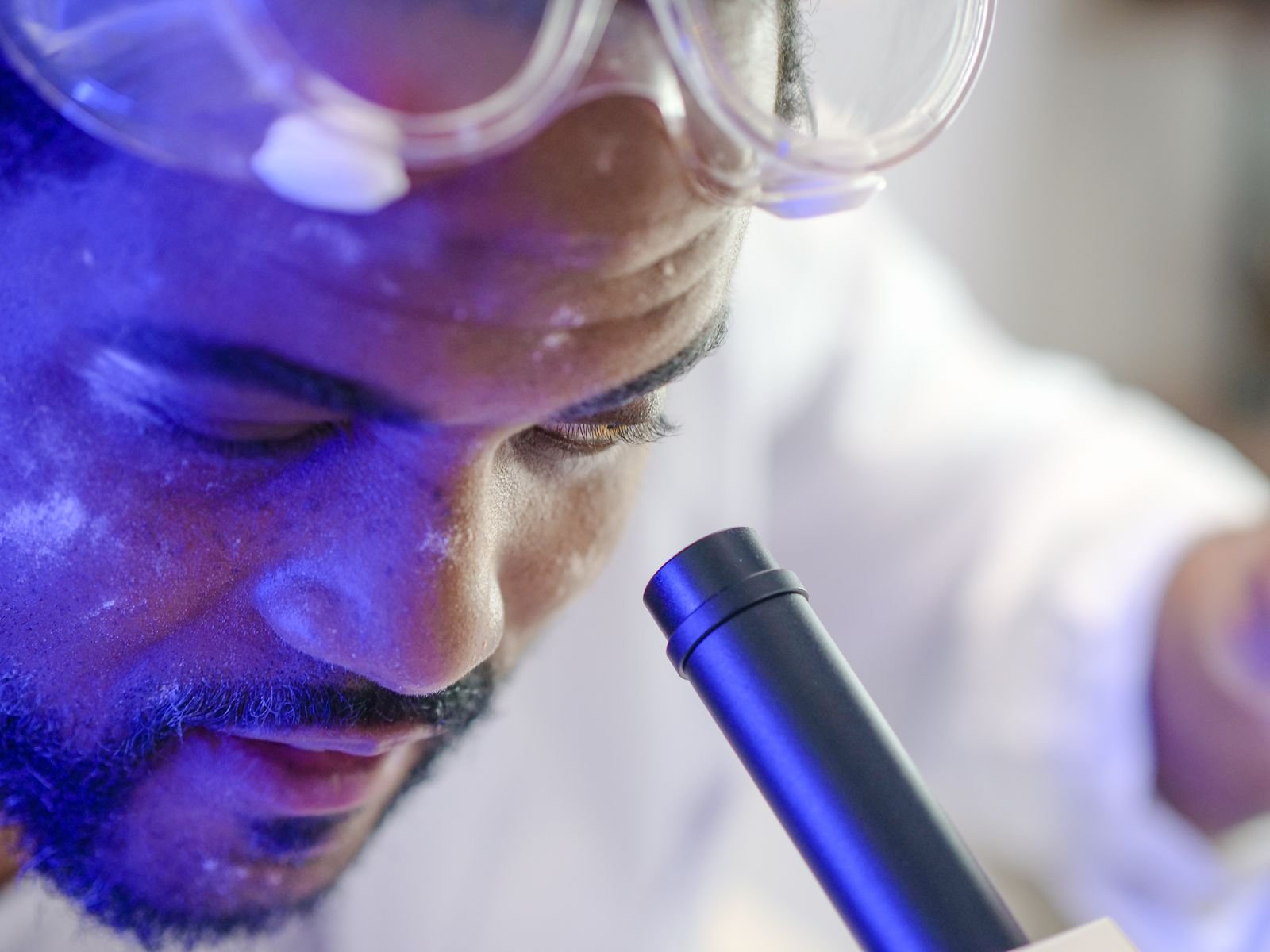
No probe. No X-ray. No sharp metal explorer walking the gumline, millimetre by millimetre. Instead, a clinician points a handheld thermal camera at your open mouth, and inflamed tissue glows back about two degrees warmer than healthy tissue. This is the promise of thermal imaging for gum disease — reading inflammation as surface heat instead of probing for it. Feed those heat patterns to a machine-learning model and, in a new study, it told advanced gum disease from a healthy mouth 98% of the time. It sounds like the end of the dreaded periodontal probe. The catch is hiding in what the camera was never asked to see.
The 30-Second Version
- A team in India used a consumer-grade thermal camera plus machine learning to separate healthy gums from advanced periodontitis using heat patterns alone — no probing, no radiation.
- The best model (AdaBoost) reached 98.15% accuracy and an ROC-AUC of 0.998, with diseased gums running roughly 2 °C warmer across every view.
- It was a careful pilot: 108 people, a single centre, and leak-resistant nested cross-validation.
- The honest caveat: it only compared healthy versus severe (Stage III/IV). The early and moderate stages — exactly where screening earns its keep — were never tested, and no outside clinic has checked the result.
It sounds revolutionary: a radiation-free, no-contact way to flag gum disease in seconds. But the number has to be read against the test behind it. In Healthcare, Ratna Bhaskar Pallipamula and colleagues at SRM University-AP and the Sibar Institute of Dental Sciences photographed 108 mouths with an infrared camera, extracted 29 temperature features per person, and pitted eleven machine-learning classifiers against each other. AdaBoost won with 98.15% accuracy. The question worth asking is not “how high did it score?” but “what, exactly, did it have to tell apart?”
The study, in one glance
This was a single-centre pilot. The researchers recruited 54 periodontally healthy adults and 54 with clinically diagnosed advanced periodontitis (Stage III or IV under the 2017 World Workshop system), all aged 35–70 with at least 20 natural teeth, and excluded anyone with diabetes, hypertension, autoimmune or thyroid disease, recent infection, antibiotics, tobacco or alcohol use — conditions that can themselves shift tissue temperature. Each person was imaged with a FLIR E6 Pro thermal camera (240×180 resolution, sensitive to 0.05 °C) in a temperature-controlled room, from five standard views, for 540 thermal images in total. From those, the team derived 29 numerical features — mean temperatures, spreads, ratios, gradients and an asymmetry index — and used only the thermal data as input, holding the clinical measurements out of the model. Performance was judged with participant-level nested five-fold cross-validation, which keeps any one person’s images from appearing in both training and test folds.
Reading gum disease without a single poke
The appeal is real, and the biology behind it is sound. Periodontal inflammation increases local blood flow, and more blood flow means a warmer gingival surface — so a camera that reads temperature is, in principle, reading inflammation. That is a genuinely attractive idea, because today’s gold-standard tools are unpleasant or risky in their own ways: the periodontal probe can make inflamed gums bleed, and repeated radiographs carry a small radiation cost. A non-contact, radiation-free snapshot sidesteps both. And the signal here was not subtle or confined to one spot — diseased gums ran warmer across all five views, by roughly 1.98 to 2.40 °C, and even a single view reached a respectable AUC of around 0.86 to 0.90. The method is careful, too: nested cross-validation and participant-level splits are the right way to keep a model from quietly memorising its own test answers.
But was the test too easy?
Here is the number that reframes the headline. The study compared healthy mouths against Stage III and IV periodontitis — the most advanced, most destructive form of the disease. It never included Stage I or Stage II. So the model was asked to distinguish two groups that a dentist can often tell apart across the room: pristine gums versus gums with deep pockets, bleeding and bone loss. Scoring 98% on that contrast is impressive engineering, but it is the easy comparison. Screening is valuable precisely when disease is early and ambiguous — the moderate, in-between cases the camera was never shown.
⚠ An “easy” binary inflates the score
Healthy versus Stage III/IV is a wide gap. The authors are explicit that this “restricted disease spectrum may result in more favourable discrimination than would be observed in a general clinical population.” A 98% here is not a promise of 98% in the grey zone where screening actually matters.
It’s a metal detector that beeps flawlessly on a cannonball and on bare sand. Impressive — but you bought it to find the coins in between.
The fine print deepens the point. The two groups also differed significantly in age, which can shift tissue temperature on its own — a confounder the authors flag honestly. Thermal regions were marked by hand with no formal inter-observer reliability check, and the camera was never blackbody-calibrated. Most important of all, every number is internal validation: no second clinic, no second camera, no different population has reproduced it.
Can you actually trust a 0.998 AUC?
Counterintuitively, an AUC of 0.998 on 108 people should raise an eyebrow as much as a cheer. Near-perfect separation on a small, single-centre dataset usually signals a task that was easily separable — not a tool ready for the clinic. The researchers resist the hype themselves: they call the work “exploratory,” decline to claim any model is statistically better than another, and write plainly that “independent external validation is required to establish generalizability.” That restraint is the most trustworthy thing in the paper. Worth knowing, too: the underlying system is the subject of a pending Indian patent held by the university.
Who could this help — and who could it mislead?
Gum disease is a global problem; the study cites an estimated 1.067 billion people living with severe periodontitis worldwide. A cheap, non-contact, radiation-free camera is exactly the kind of tool that could extend screening into community clinics where probing and radiography are scarce. That is the equity upside. But the same openness is a risk. The study ran in a controlled 23–25 °C room, with participants acclimatised and confounders like diabetes, recent meals and physical activity excluded — none of which real mouths guarantee. Surface temperature is sensitive to ambient conditions, recent food and drink, and physiology that varies across people, so a tool validated in a quiet, filtered setting can behave very differently in a busy, warm one. A confidently wrong “all clear” is worse than no screen at all.
What this means for you
If you’re a patient
This is promising research, not a product you can ask for. No thermal camera is going to replace the probe and X-ray that diagnose and stage gum disease today. If your gums bleed, recede or feel loose, that is still a reason to see a dentist — not to wait for a heat scan.
If you’re a clinician
Read it as a proof of concept for a complementary flag, not a diagnostic. The design can’t tell you how it performs on early disease, across devices, or in a warm operatory. Watch for the external, multi-centre validation the authors call for before it means anything chairside.
The bottom line
Infrared thermography can clearly “see” the heat of advanced gum disease, and the engineering here is clean. But a near-perfect score on the easiest possible comparison is a starting line, not a finish. The honest read: a thermal camera may one day earn a place as a non-contact screening aid — an assistant that points you toward a closer look, never an oracle that replaces it.
Frequently asked questions
Can a thermal camera really detect gum disease?
In this study, yes — but only in a narrow sense. A machine-learning model reading infrared images separated healthy mouths from advanced (Stage III/IV) periodontitis with 98.15% accuracy, because diseased gums ran about 2 °C warmer. It was never tested on early or moderate disease, and it has not been validated outside the single clinic that built it, so it is a research signal, not a clinical test.
Does this mean no more probing or dental X-rays?
No. Probing depth, attachment loss and radiographs are still how periodontitis is diagnosed and staged. The thermal method is being explored as a possible non-contact, radiation-free complement to those tools — not a replacement for them.
Why is a 98% accuracy result not as strong as it sounds?
Because the model only had to tell obviously healthy gums from the most severe disease — the easy extremes. It skipped the early and moderate stages where screening is most useful and hardest. High accuracy on an easy comparison does not predict high accuracy in the ambiguous middle.
What would make this trustworthy enough to use?
External, multi-centre validation on a full spectrum of disease (including Stage I and II), across different cameras and real-world room conditions, with calibrated devices and tested reproducibility. The authors say as much themselves.
Is thermal imaging safe?
Yes — infrared thermography is passive and non-contact. It simply measures the heat a surface gives off, with no radiation and no physical contact, which is a large part of why it is appealing for screening.
Source & author credit
This article interprets, and does not reproduce, the following peer-reviewed study. All figures are the authors’ original findings.
Pallipamula RB, Donepudi SP, Jahanavi K, Brindha A, Katuri KK, Dhulipalla R, Sunitha KA. An Exploratory Study Investigating the Intraoral Thermal Features in Advanced Periodontitis Using Machine Learning Techniques. Healthcare (Basel). 2026;14(21):3749. DOI: 10.3390/healthcare14213749
ORCID — Pallipamula 0009-0000-6320-8181 · Donepudi 0009-0007-0815-6594 · Jahanavi 0009-0003-6079-5650 · Brindha 0000-0003-2722-0057 · Katuri 0000-0003-2002-4352 · Dhulipalla 0000-0003-4103-455X · Sunitha 0000-0003-4232-8787
Published open access under a Creative Commons Attribution (CC BY 4.0) licence. Decadentry is an independent educational publication and is not affiliated with the study’s authors. For how we select and verify studies, see our editorial standards, read more in our periodontology research hub, and compare this with our look at whether AI can forecast gum disease.
Leave a Reply