

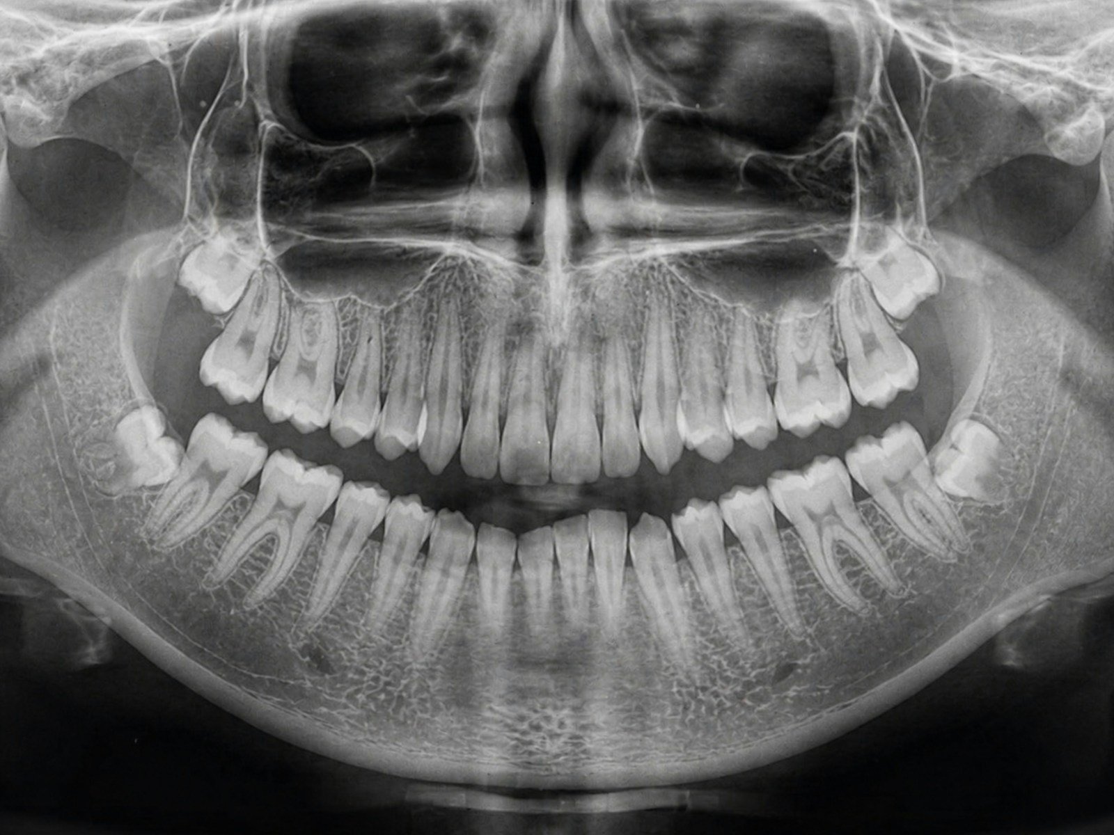
A radiolucent jaw lesion is easy to miss precisely because it is quiet. On a panoramic X-ray taken before a crown or a wisdom-tooth extraction, a cyst or tumour shows up as a soft grey shadow in the bone — no pain, no swelling, nothing the patient can feel. Catch it early and it is often a minor procedure; miss it for a few years and it can hollow out the jaw. That gap between “seen” and “missed” is exactly where AI jaw lesion detection is being asked to help.
The 30-Second Version
- A 2026 study trained four deep-learning object detectors — YOLOv5, YOLOv8, YOLO26 and Faster R-CNN — to spot radiolucent jaw lesions (cysts, tumours and other dark patches) on 600 panoramic X-rays.
- The models agreed on roughly one thing: they were good but not great. The best F1 score was about 70%, and recall ranged from 59% to 76%.
- They differed in style: YOLOv8 was the most cautious (79% precision, but it caught only 59% of lesions), while YOLOv5 and Faster R-CNN caught more (76%) at the cost of more false alarms.
- Strict localisation was weak — a mean average precision of just 30–32% at tight overlap — and it is one dataset, 60 test images, no external validation.
It sounds like the ideal second pair of eyes. But the claim deserves scrutiny. In the Journal of Stomatology, Oral and Maxillofacial Surgery (2026), Emre Yapıcıoğlu and Kamil Serkan Ağaçayak compared four popular detection networks on the same set of 600 annotated panoramic radiographs, asking a deceptively simple question: which architecture is best at drawing a box around a jaw lesion — and is any of them good enough to trust? The answer is more interesting than a single winner.
The study, in one glance
The researchers collected 600 panoramic radiographs that each showed a radiolucent jaw lesion, anonymised them, and had the lesions outlined with rectangular bounding boxes. The images were split into training (80%), validation (10%) and a held-out internal test set (10%, about 60 images). Data augmentation applied only to the training set expanded it from 600 to 1,142 images. Four object-detection models — YOLOv5, YOLOv8, YOLO26 and the two-stage Faster R-CNN — were then trained and tested on identical data splits, and scored on precision, recall, F1 and mean average precision (mAP).
The promise: AI jaw lesion detection as a tireless second reader
Here is why AI jaw lesion detection is worth taking seriously. A general dentist taking a panoramic image is usually focused on teeth, roots and the odd impacted molar — not systematically hunting the quiet grey shadows of a cyst or tumour in the body of the mandible. Those lesions are often found by accident, if at all. A model that scans every panoramic and quietly boxes the suspicious areas is the kind of opportunistic safety net that turns a routine image into an early warning, the same logic behind using a dental X-ray to flag calcified neck arteries. In this study the models reached F1 scores around 70% with recall as high as 76% — pointing, for the lesions present, at roughly three out of four. Against a human baseline of “whatever the busy clinician happens to notice,” that is a meaningful lift.
So why did the best model still miss a quarter of lesions?
Because “good” and “safe enough to rely on” are different thresholds. The headline recall of 76% means that even the better detectors missed about one lesion in four — and YOLOv8, the most cautious model, missed 41% of them while being right 79% of the time when it did flag something. That is the central tension the authors highlight: the four networks posted similar F1 scores but struck very different bargains between precision and recall. There is no free lunch here. Tune a model to miss fewer lesions and it flags more healthy bone; tune it to be sure of its alerts and it stays silent on real disease.
⚠ A box is not a diagnosis
Detecting a lesion is not the same as identifying it. These models drew boxes around radiolucencies; they did not say whether a shadow is a harmless cyst, an aggressive tumour or an artefact. Any flagged area still needs a clinician — and usually a biopsy or CBCT — to say what it actually is.
It is less a diagnostician than a metal detector on a beach: it beeps where something might be buried, but someone still has to dig, and it beeps for bottle caps too.
The localisation numbers sharpen the point. At a lenient overlap threshold the best mAP reached 70% (Faster R-CNN), but at the strict mAP@0.5:0.95 threshold — which demands the predicted box sit tightly on the true lesion — every model collapsed to 30–32%. In plain terms, the detectors were far better at saying “something is roughly here” than at tracing exactly where a lesion begins and ends, a limitation the authors attribute partly to scanning the entire panoramic field, where normal anatomy offers endless distractions.
Can you trust a number from a single dataset?
This is where enthusiasm should slow down. All 600 images came from one source and were split internally; the test set was just 60 images; and the paper reports no external validation on radiographs from other clinics, scanners or populations. Deep-learning detectors are notorious for looking brilliant on their home data and stumbling on the first unfamiliar machine. The authors frame this as a comparative, moderate-performance study — a map of how four architectures behave, not a finished tool. They add a practical nudge: because two-stage detectors like Faster R-CNN are slower, the faster YOLO models may be the more realistic choice for chairside support when performance is comparable.
Who benefits — and who never gets the second look?
An AI second reader only ever helps the images it sees. Jaw lesions can be most consequential in people with the least access to regular dental imaging, yet a detector bolted onto a modern practice’s panoramic software reaches exactly the patients already in the chair. And a model trained on one population may quietly underperform on jaws it was never shown. Without external validation across diverse settings, a tool that promises to catch more could, in practice, catch more only for the already-served.
What this means for you
If you’re a patient
Nothing here changes your next visit, and there is no tool to request. If a dentist mentions a shadow on your panoramic X-ray, treat it as a reason for a proper look — a CBCT scan or specialist referral — not as a verdict. The value of AI here is helping a clinician not overlook something, not replacing their judgement.
If you’re a clinician
Read these as comparative benchmarks, not a green light. Recall in the 70s with strict-localisation mAP near 30% means a model can miss lesions and box them loosely. If you trial detection software, confirm every flag yourself, keep your own reading discipline, and wait for externally validated, multi-centre results before leaning on the numbers.
The bottom line
Four modern detectors can all find jaw lesions on a panoramic X-ray about as well as each other — which is to say, usefully but imperfectly, catching most lesions while missing a stubborn minority and localising them only loosely. That makes this class of tool a promising assistant that nudges a busy clinician to look again, not an oracle that reads the film for them. The honest headline is not “AI finds jaw lesions” but “AI finds most jaw lesions, on one hospital’s images, if a human checks its work.”
Frequently asked questions
Can AI detect jaw lesions on a dental X-ray?
Yes, to a degree. In a 2026 study, deep-learning object-detection models located radiolucent jaw lesions on panoramic radiographs with F1 scores around 70% and recall up to 76%. But they detect and box lesions rather than diagnose them, and the best model still missed about a quarter of lesions, so they work as an assistant to a clinician, not a replacement.
Which AI model was best at finding jaw lesions?
No single model dominated. YOLOv8 had the highest precision (79%) but the lowest recall (59%), while YOLOv5 and Faster R-CNN caught the most lesions (76% recall) with more false alarms. F1 scores were similar across all four, and Faster R-CNN had the best overall mAP@0.5 (70%).
Does the AI know whether a lesion is a cyst or a tumour?
No. The models only drew bounding boxes around radiolucent areas; they did not classify a lesion as a cyst, tumour or artefact. Determining what a shadow actually is still requires clinical assessment and often a CBCT scan or biopsy.
Is this tool ready for everyday dental practice?
Not yet. The study used 600 images from a single source with only about 60 test images and no external validation on other clinics or scanners. Strict localisation accuracy was also low (around 30%). It is a research comparison of architectures, not an approved, broadly validated clinical product.
Source & author credit
This article interprets, and does not reproduce, the following peer-reviewed study. All figures are the authors’ original findings.
Yapıcıoğlu E, Ağaçayak KS. Deep learning-based detection of radiolucent jaw lesions on panoramic radiographs: A comparative study. Journal of Stomatology, Oral and Maxillofacial Surgery. 2026;127(6):103018. DOI: 10.1016/j.jormas.2026.103018
ORCID — Yapıcıoğlu 0009-0003-6215-5542 · Ağaçayak 0000-0002-6168-1802
© 2026 Elsevier Masson SAS. All rights reserved; this article summarises and does not reproduce the copyrighted original. Decadentry articles are checked against their primary source per our editorial standards, and sit within our Diagnostics & Imaging research hub. Decadentry is an independent educational publication and is not affiliated with the study’s authors.
Leave a Reply